Signalment:
Female rabbit (
Orytolagus cuniculus), adult (unknown age)The rabbit was found outdoors with a mammary
mass (sub-Q mass about 5-6 cm diameter) in the caudal
ventral abdomen that turned out to be a malignant hair
follicle tumor. The uterus was removed at a later date
because an abdominal mass was palpated and to spay the
rabbit.
Gross Description:
Firm pale masses, thickened friable
mucosa, and multiple mucosal cysts.
Histopathologic Description:
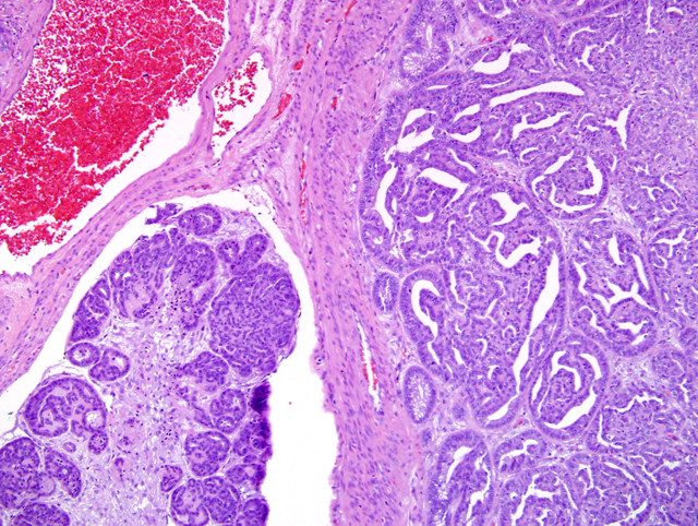
Histologically,
there are two distinctly different masses present within
the same section adjacent to the mesometrial insertion;
the first located in the endometrium, the second located
within the myometrium. The masses are located adjacent
to one another and in some areas are in direct contact. The
endometrial mass is multilobulated and well demarcated
but unencapsulated. It is composed of thick papillary
projections expanding into the uterine lumen, as well as
lobules of densely packed tubular and glandular structures
infiltrating into the subjacent myometrium (
Fig. 2-1).
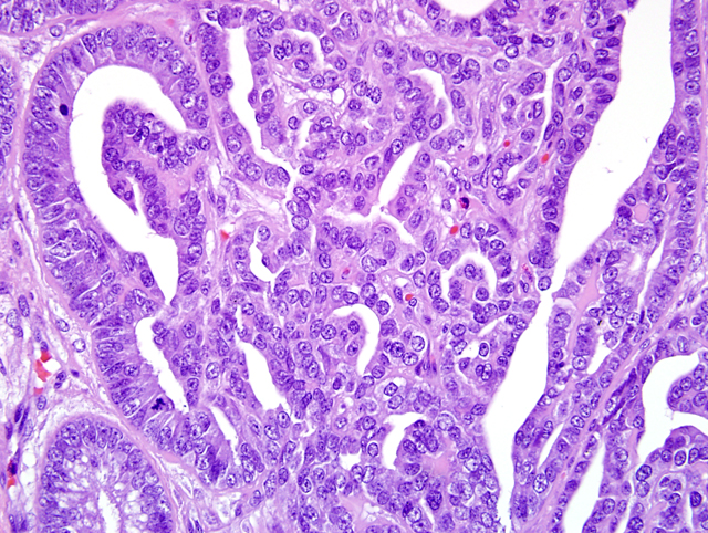
Within the papillary projections and lobules, cuboidal
to columnar neoplastic epithelial cells form a single,
albeit crowded, layer supported by variable amounts of
loose vascular collagenous stroma (
Fig. 2-2). Lobules
are separated by moderate amounts of loose collagenous
or myxomatous stroma, while within lobules, cells are
supported by scant stroma. The neoplastic population
has moderate anisocytosis and anisokaryosis. There are
1-3 mitotic figures per 400X field. Minimal amounts of
sloughed cells, inflammation and necrosis are present
within the mass. In some sections there are lobules of
neoplastic cells within uterine lymphatic vessels covered
by a layer of endothelium. Microscopic features of this
mass are consistent with an adenocarcinoma.
2
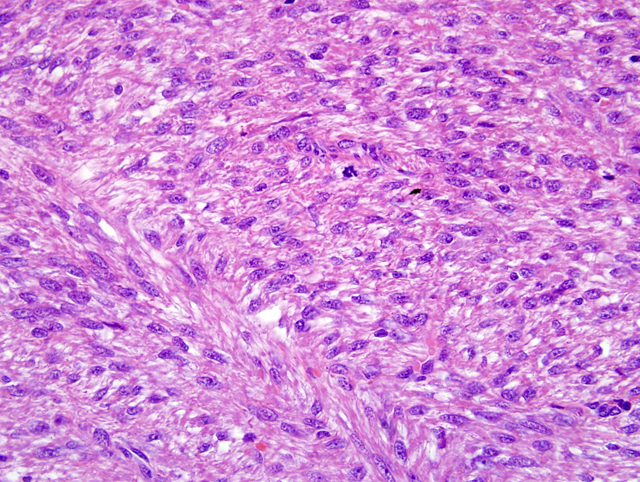
Microscopically, the second mass, located in the
myometrium, is multilobulated, unencapsulated, poorly
demarcated in some areas and infiltrative. It is composed
of densely cellular, broad interlacing fascicles of medium
sized spindloid cells (
Fig. 2-3). These neoplastic cells
have indistinct cytoplasmic borders, moderate amounts
of vacuolated or fibrillar eosinophilic cytoplasm, a bluntended
oval nucleus with finely granulated chromatin
and a single eosinophilic nucleolus. In general, there
is slight anisocytosis and anisokaryosis, however, rare
karyomegalic and multinucleated cells are present. Mitotic
figures are uncommon (1 per ten 400X fields). There are
several large areas of necrosis with hemorrhage present
within the mass (not present in all sections, Images 1 and
2). Although the mitotic rate is low and the neoplastic cells
are well-differentiated, the presence of areas of necrosis is
consistent with a diagnosis of leiomyosarcoma rather than
leiomyoma.
2
Morphologic Diagnosis:
Uterus: Adenocarcinoma
Uterus: Leiomyosarcoma
Condition:
Uterine adenocarcinoma, leiomyoma
Contributor Comment:
In contrast to other
domestic species where uterine adenocarcinomas are a rare
occurrence, the incidence of uterine adenocarcinomas has
been reported to be up to 79% in female rabbits over the
age of five years, with variation in breed predisposition.
In fact, this tumor type is the most common tumor of
Orytolagus cuniculus overall.
6 Uterine adenocarcinomas
in rabbits are often multicentric and affect both uterine
horns.
1Most are slow growing and present with bloody
discharge and/or decreased fertility in breeding animals.
5,6
The pathogenesis of these tumors is unclear and appears
to be different from those occurring in humans, although
there is controversy over this subject in the literature.
Uterine leiomyosarcomas are much less common than
adenocarcinomas with reported incidences of 1% in
aging Dutch rabbits, and 2% in rabbits presenting to
veterinarians with uterine disorders.
1,5 The submitted case
represents a simultaneous occurrence of two spontaneous
uterine tumors in a female rabbit. Recently a similar
case of concurrent adenocarcinoma and leiomyoma in an
individual animal was reported by Kurotaki et al.
4 The
case report describes two endometrial adenocarcinomas,
one of which was closely associated with a myometrial
leiomyoma. These cases are similar in the presence of
both primary epithelial and mesenchymal tumors within
the uterus, with a close physical association between the
two. They are different in that the smooth muscle tumor
is malignant in the submitted case.
JPC Diagnosis:
Uterus: Adenocarcinoma
Uterus: Leiomyosarcoma
Conference Comment:
The cow is the only
other domesticated animal that commonly gets uterine
adenocarcinomas. This tumor is often seen in abbatoirs
and grossly appears as single or multiple firm masses in the
wall of the uterus.
3 These tumors are often umbilicated and
are known to metastasize to the internal iliac lymph nodes
and lungs.
3 Histologically, this tumor often causes an
intense fibrous response with numerous mitoses, cellular
pleomorphism, and vascular and lymphatic invasion.
3
Other differentials for a neoplastic growth in the uterus
of domesticated animals include adenoma, fibroma,
leiomyoma, and leiomyosarcoma as mentioned by the
contributor. Adenomas are generally rare in domesticated
species and can be difficult to distinguish from focal
endometrial hyperplasia.
3 Fibromas are generally firm,
white, expansile masses that histologically appear as very
bland, sparsely cellular, densely collagenous growths.
3
Fibromas are seen most commonly in dogs and cattle, but
they are not prevalent tumors.
3 Leiomyomas are composed
of interwoven bundles of smooth muscle originating in
the myometrium. The amount of connective tissue can
be highly variable.
3 These tumors are seen in dogs, cats,
and cattle.
3
References:
1. Baba N, von Haam E: Animal Model: Spontaneous
adenocarcinoma in aged rabbits. Am J Path
68:653-656,
1972
2. Cooper BJ, Valentine BA: Tumors of Muscle.Â
In:
Tumors of Domestic Animals, ed Meuten DJ, 4th ed., 319-
363, 2002
3. Kennedy PC, Cullen JM, Edwards JF, Goldschmidt
MH, Larsen S, Munson L, Nielsen S: Histological
classification of tumors of the genital system of domestic
animals.Â
In: World Health Organization Histological
Classification of Tumors of Domestic Animals, ed.
Schulman FY, Second Series, vol. 4, pp. 32-33, 72. Armed
Forces Institute of Pathology,Washington, DC, 1999
4. Kurotaki T, Kokoshima H, Kitamori F, Kitamori
T, Tsuchitani M: A case of adenocarcinoma of the
endometrium extending into the leiomyoma of the uterus
in a rabbit. J Vet Med Sci
69:981-984, 2007
5. Saito K, Nakanishi M, Hasegawa A: Uterine disorders
diagnosed by ventrotomy in 47 rabbits. J Vet Med Sci
64(6)495-497, 2002
6. Weisbroth SH: Neoplastic Diseases.Â
In: The Biology
of the Laboratory Rabbit, eds. Manning PJ, Ringler DH,
Newcomer CE, 2nd ed., 259292, Academic Press, New
York, NY, 1994