Signalment:
Male, Coimbra's titi (
Callicebus coimbrai)This monkey was kept at the zoological
park in Belo Horizonte, Brazil. It was found on the floor in a cage, prostrated
and hypothermic. It received emergency therapy with fluids, corticosteroids,
glucose, and heating, but died soon after the initial treatment.
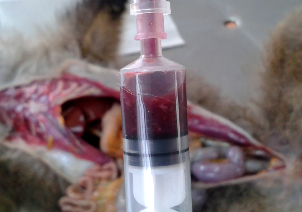
Gross Description:
Grossly, the mesenteric lymph nodes were
hemorrhagic, the colon was dilated with multifocal and moderate hemorrhage in
the serosa. The colon content was hemorrhagic with a few blood clots, and large
amounts of fibrinous exudate. There was sand in the oral cavity, esophagus, and
stomach. The heart was mildly dilated. On the surface and cut surfaces of the
liver, there was a prominent lobular pattern. Kidneys and adrenal glands were
moderately congested.
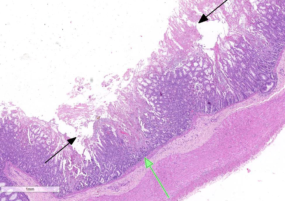
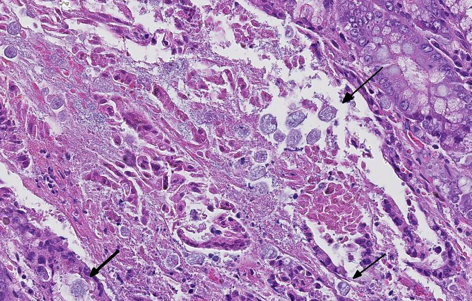
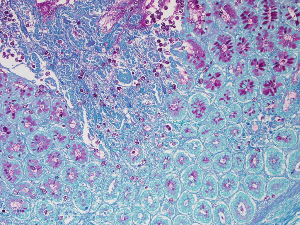
Histopathologic Description:
Multifocal to
coalescing and severe necrosis associated to multifocal and moderate hemorrhage was observed in the mucosa of
the colon. Myriads of round, 30-50 m
diameter, eosinophilic staining trophozoites were diffusely distributed in the
lumen, necrotic mucosa, crypts, and lamina propria of the colon. There was a
multifocal and mild infl-ammatory infiltrate composed of ly-mphocytes,
histiocytes and neutrophils in mucosa and submucosa. Some epithelial cells of
the colonic crypts had numerous circular, intensely basophilic cytoplasmic
structures, which were interpreted as apoptotic bodies.
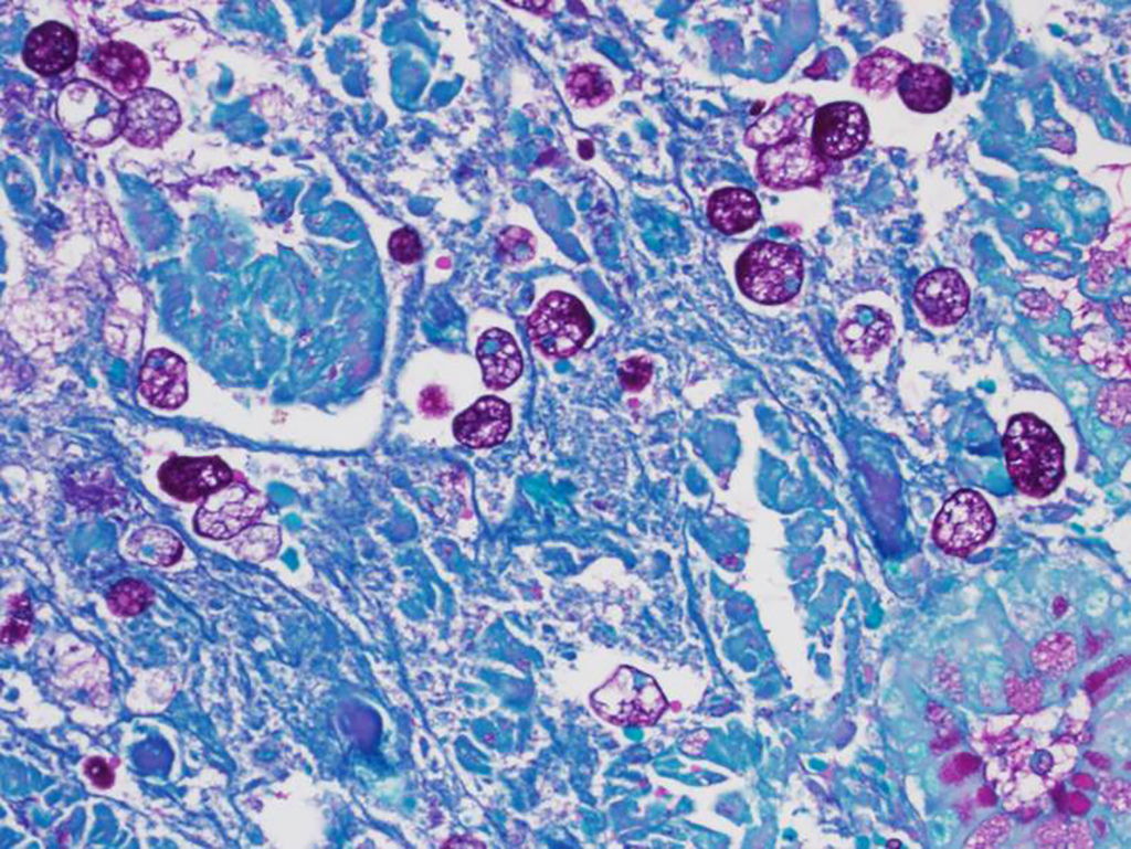
Morphologic Diagnosis:
Large
intestine, colon: Colitis, necrotizing, hemorrhagic, acute, multifocal to co-alescing,
severe with numerous protozoal organisms consistent with
Entamoeba
histolytica.
Lab Results:
None.
Condition:
Entamoeba histolytica, titi monkey
Contributor Comment:
Entamoeba
histolytica
is a protozoan parasite capable of invading the intestinal mucosa. It affects
other organs, mainly the liver, causing amebiasis.7 Entamoeba
dispar is mor-phologically similar. It also colonizes the human gut,
however it has no invasive potential.1 Both species are found of
non-human primates, such as monkeys, orangutans, and baboons.5
Most asymptomatic
infections found worldwide are now at-tributed to
Entamoeba dispar,
because it is non-invasive.
E. dispar is distinct but closely related to
E. histolytica. This non-invasive species has im-plications for
understanding the epidemiology of amebiasis.
3
In humans,
amebiasis is more common in developing countries. Bad conditions such as
overcrowding, poor education, co-ntaminated water, and poor sanitation favor
fecal-oral transmission of amoebas. This disease results in 70 thousand deaths
annually. Amebiasis is considered the fourth cause of death due to protozoa.
11A recent study revealed a high prevalence of
E. histolytica in
long-tailed macaques in the Philippines.
8 Captive monkeys infected
with
E. histolytica is a concern not only for the animal health risk,
but also due to the zoonotic nature of the disease.
4
The trophozoite,
which is the motile form of
E. histolytica, lives in lumen of crypts in
large intestine, where it multiplies and differentiates into cyst (resistant
form responsible for transmission). Cysts are excreted in stools, and may be
ingested by a new host through contaminated food or water. Upon ingestion of
infective cysts, parasites are released in terminal ileum, with each emerging
quadrinucleate trophozoite giving rise to eight uninucleated trophozoites.
Trophozoites may invade the colonic mucosa and cause dysentery.
3
There
are some molecules produced by
E. histolytica that are related to lysis
of the colonic mucosa: adhesins, amoebapores, and proteases. The parasite
attachment to colonic mucus blanket is due to a multifunctional adherent
lectin, preventing elimination in intestinal stream. Lectins are involved in
signaling cytolysis and in blocking the deposition of the injurious membrane
attack complex of complement. It maybe also participates in anchorage of the
ameba to proteoglycans, during invasion process.
3 Peptides of
E.
histolytica named am-oebapores destroy phagocytosed bacteria from the
microbiota that serve as the main nutrient source for the parasite. Whether
amoebapores play a role in the cytolytic event has not yet been proven.
Proteases produced by the parasite can degrade the extracellular matrix during
invasion and contribute in the lysis of target cells.
3
JPC Diagnosis:
Colon: Colitis, necrotizing, acute, multifocally extensive, marked with crypt abscesses, goblet
cell loss and numerous amoebic trophozoites.
Conference Comment:
E.
histolytica
colonizes the large intestine resulting in colitis and diarrhea,
and in some cases, may result in liver abscesses. E. histolytica has
two morphogenetic forms: the trophozoite and infectious cyst form. Ingestion
of viable cysts initiates infection and subsequent steps are discussed above. The
initial attachment to mucosal enterocytes by trophozoites, mediated through
adhesins and lectins, appears to be important in the initial pathogenesis and
apparently plays a role in cytotoxic activity.6 The host
inflammatory response contributes to tissue damage and may help facilitate
infection. Important cytokines involved in the host response include IL-8,
IL-6 and IL-1?; genes involved in cell proliferation also participate in the
response to infection. E. histolytica destroys host tissue and commonly
causes enterocyte apoptosis; trophozoites may ingest host cells following their
death. The mechanisms leading to host cell apoptosis are not completely
understood, but may involve production of reactive oxygen species and oxidative
stress. Other mechanisms which may be involved in cell death include amoebic
trogocytosis, where the parasite ingests fragments of host cell, resulting in
increases in intracellular calcium and cell death. 2 Other parasite
factors which are
involved in the pathogenesis include production of prostaglandin E2, which
increases sodium ion permeability through tight junctions, as well as secretion
of cysteine proteases which digest matrix components such as gelatin, collagen
type I and fibronectin. Epithelial barrier disruption also plays an important
role in infection.2 E. histolytica
trophozoites normally remain in the colonic lumen. However, in some cases, the
trophozoites invade the mucosa as well as mural blood vessels and lymphatics,
and eventually infect the liver. Ulcerative gastritis secondary to E.
histolytica infection has also been described in primates that have a
sacculated stomach, which is adapted for leaf eating and fermentation.
Similarly, E. histolytica-associated gastritis may also occur in
macropods (kangaroos and wallabies), which likewise have a sacculated stomach.
9 Trophozoites are observed in the mucosa and gastric glands, but may
also invade to the level of the submucosa, including vessels and
lymphatics.
E. histolytica may
also cause necrotizing and ulcerative colitis in dogs and cats (likely acquired
from a human source) although less commonly than in primates. In cats, E.
histolytica infection has been associated with severe necrosis of the colon
and cecum.9,10 It is apparently uncommon for infected dogs to shed
infectious cysts.
References:
1.
Diamond LS, Clark CG. A redescription of Entamoeba histolytica
Schaudinn, 1903 (emended Walker, 1911) separating it from Entamoeba dispar
Brumpt, 1925. J Eukaryot Microbiol. 1993; 40: 340–344.
2. Di
Genova BM, Tonelli RR. Infection strategies of intestinal parasite pathogens
and host cell responses. Front Microbiol. 2016; 7:256. doi:
10.3389/fmicb.
3.
Espinosa-Cantellano M, Martínez-Palomo A. Pathogenesis of Intestinal Amebiasis:
From molecules to disease. Clin Microbiol Rev. 2000; 13(2):318.
4.
Feng M, Cai J, Min X, Yongfeng F, Xu Q, Tachibana H, Cheng X. Prevalence and
genetic diversity of Entamoeba species infecting macaques in southwest China. Parasitol
Res. 2013; 112:1529–1536.
5.
Feng M, Yang B, Yang L, Fu YF, Zhuang YJ, Liang LG, Xu Q, Cheng XJ, Tachibana
H. High prevalence of Entamoeba-infections in captive long-tailed macaques in
China. Parasitol Res. 2011; 109:1093–1097.
6. García MA, Gutiérrez-Kobeh L, López Vancell R. Entamoeba
histolytica: Adhesins and Lectins in the trophozoite surface. Molecules.
2015; 20:2802-2815.
7.
Martínez-Palomo A, Espinosa-Cantellano M. Amoebiasis: new understanding and new
goals. Parasitol Today. 1997; 14:1–3.
8. Rivera WL, Yason JA, Adao DE. Entamoeba histolytica and E.
dispar infections in captive macaques (Macaca fascicularis) in the
Philippines. Primates. 2010; 51:69–74.
9.
Stedman NL, Munday JS, Esbeck R, Visvesvara GS. Gastric amebiasis due to Entamoeba
histolytica in a Dama Wallaby (Macropus eugenii). Vet Pathol.
2003;40:340-342.
10. Uzal FA, Plattner BL, Hostetter JM. Alimentary system. In: Maxie MG, ed. Jubb, Kennedy, and
Palmer's Pathology of Domestic Animals. 6th ed. Vol 2. St. Louis, MO:
Elsevier; 2016:98-99.
11.
World Health Organization. 1998. The World Health Report 1998. Life in the 21st
century: a vision for all. World Health Organization: Geneva, Switzerland.