Signalment:
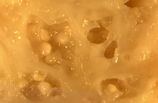
Gross Description:
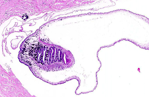
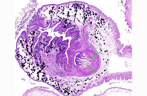
Histopathologic Description:
Morphologic Diagnosis:
1. Skeletal muscle: Cysticercosis with eosinophilic, granulomatous myositis, fibrosis, and myodegeneration.
2. Skeletal muscle: Sarcocytosis
Condition:
Contributor Comment:
Feral woodchucks may bear heavy cysticerci burdens that may be particularly prominent after emergence from hibernation.(2) Laboratory woodchucks may also develop cysticercosis, especially if the animals are of wild-caught origin.(1,5)
The species of tapeworm affecting this woodchuck is not definitively known. Previously reported cases of woodchuck cysticercosis were consistent with T. crassiceps larvae; the fox was considered the likely definitive host in these cases. (1-3,5) Morphologic features of this parasite are consistent with T. crassiceps and include an anterior scolex with four suckers, an apical rostellum that contains large and small rostellar hooks, and a posterior bladder.(1,4,5)
The inflammatory response in this case is relatively mild and composed of a mixed population of inflammatory cells. Inflammation is more intense in areas of cyst rupture. Previous reports of the inflammatory reaction woodchuck cysticercosis describe a similarly mild response with more prominent lymphocytic infiltrates and a similar variability in the degree of reactive fibrosis.(1)
JPC Diagnosis:
Conference Comment:
There are two orders in the phylum Platyhelminthes which comprise tapeworms.(4) The order of pseudophyllideans grows into much larger adults, such as Diphyllobothrium spp., and never shed their proglottids. This is contrast to the order of cyclophyllideans, as observed in this case, which shed gravid proglottids each containing thousands of infectious eggs. The cyclophyllideans are more readily transmissible and as a result, are the most significant cause of CNS disease in people in South America.(6) When the eggs are ingested, the larvae migrate into various tissues resulting in significant pathology. Conference participants speculated on what drove these larvae to all migrate to the same location in this woodchuck.
We agree with the contributor that, although T. cracisseps is the most often reported and most likely species in this case, the presence of an anterior scolex with four suckers and a posterior bladder is only indicative of the type of larval cestodes known as a cysticercus and cannot be differentiated further. However, some references indicate the possibility of species identification based on the length of small and large hooks in the rostellum.(3) Other groups of larval cestodes include the cysticercoids which have a tiny bladder and a scolex surrounded by parenchymous arms, coenurus which has more than one scolex, and hydatid cysts with a bladder and numerous small protoscolices each with a scolex and suckers. Solid-bodied cestodes are plerocercoids (lack suckers) or tetrathyridium (has suckers).(4)
References:
1. Anderson WI, Scott DW, Hornbuckle WE, King JM, Tennant BC: Taenia crassiceps infection in the woodchuck: a retrospective study of 13 cases. Vet Dermatol. 1: 85-92, 1990.
2. Beaudoin RL, Davis DE, Murrell KD: Antibodies to larval Taenia crassiceps in hibernating woodchucks, Marmota monax. Exp Parasitol. 24(1): 42-6, 1969.
3. Brojer CM, Peregrine AS, Barker IK, Carreno RA, Post C: Cerebral Cysticercosis in a Woodchuck (Marmota monax). Journal of Wildlife Diseases. 38(3): 621624, 2002.
4. Gardiner CH and Poynton SL: Morphologic characteristics of cestodes in tissue section. In: An atlas of metazoan parasites in animal tissues: American Registry of Pathology, Washington, D.C. 199, 50-55.
5. Shiga J, Aoymana H, Yamamoto K, Imai S, Saeki H, Sasaki N, Koshimizu K: A case report of cysticercosis caused by Cysticercus longicollis, a larval form of Taenia crassiceps in a woodchuck (Marmota monax). Jikken Dobutsu. 36(2): 213-7, 1987.
6. http://www.cdc.gov/parasites/cysticercosis. Center for Disease Control. April 16, 2014.