WSC 22-23
Conference 5
CASE IV:
Signalment:
5 year, 2 month old female Beagle dog (Canis familiaris)
History:
A multiparous female from a breeding colony was noted to have bilaterally elevated nictitating membranes and a body temperature of 99.8 °F. No other clinical signs were noted. Two days later the dog was found moribund and was humanely euthanized. Approximately 5 years prior to this episode, the dog had been treated for respiratory disease (pneumonia).
Gross Pathology:
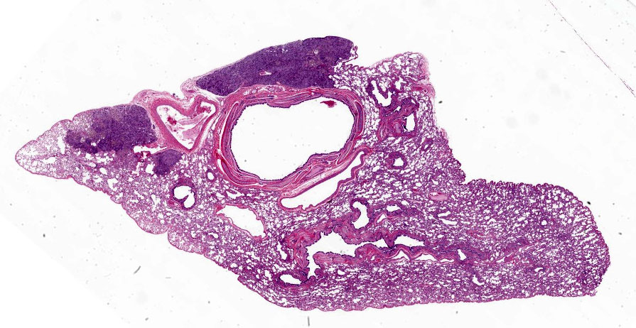
The animal was in good body condition with copious fat deposits. Caudal lung lobes contained multiple, gray brown, 0.5 - 1 cm diameter subpleural nodules. Stomach contents were dark brown with flecks. Small areas of petechiation and yellow chyme-like material were noted in the duodenal mucosa and lumen respectively. No other gross lesions were noted.
Laboratory Results:
No laboratory findings reported.
Microscopic Description:
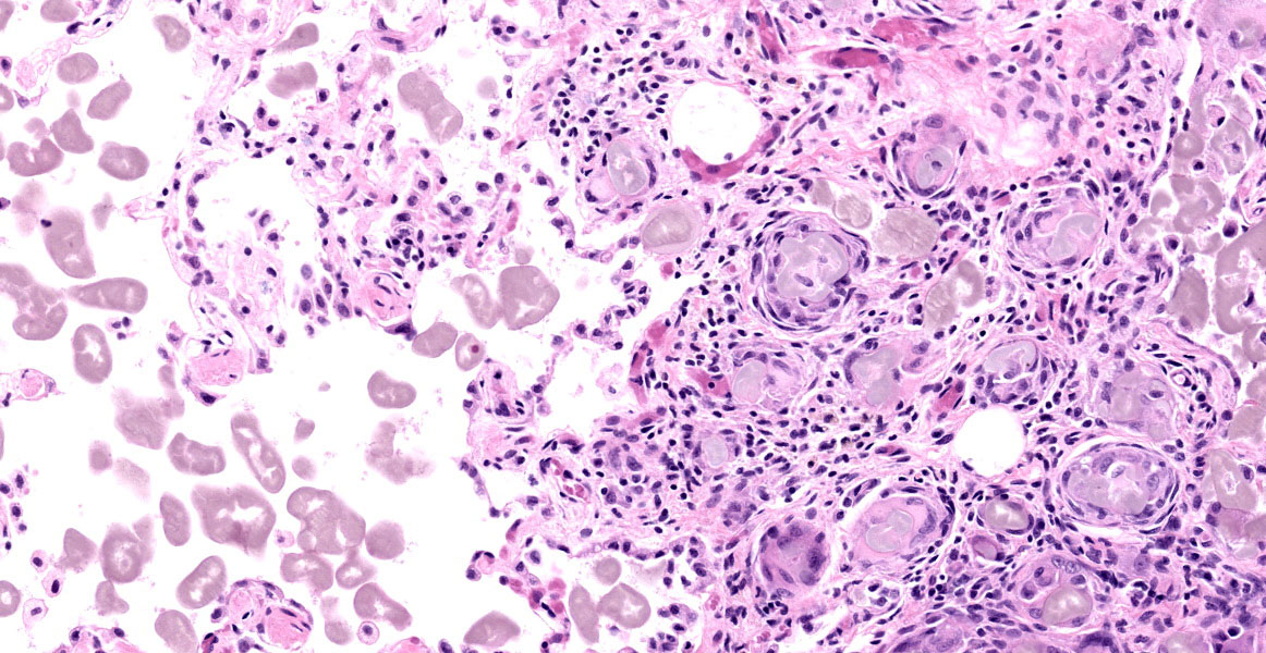
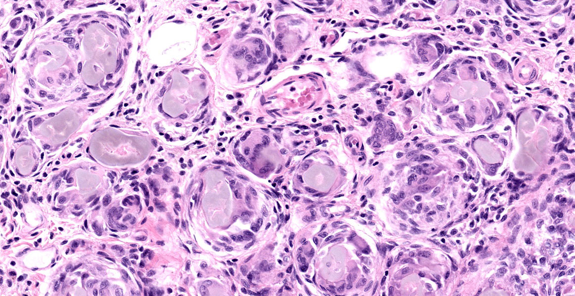
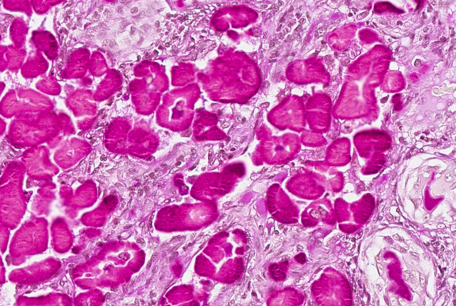
Lung: In one or more subpleural or apical foci, alveoli are filled with macrophages, multinucleate giant cells, and amorphous pale gray-brown material; small accumulations of amorphous material are often within multinucleate giant cells. Some alveoli contain only amorphous material (without inflammatory cells). Alveolar septa are thickened by fibrous connective tissue and infiltrated by varying numbers of mononuclear inflammatory cells. Amorphous material is strongly PAS positive, and exhibits birefringent peripheral radial striation under polarized light.
Contributor’s Morphologic Diagnoses:
Pneumonia, granulomatous, multifocal, chronic, with intralesional hyaline material.
Contributor’s Comment:
Pulmonary hyalinosis is considered an incidental finding in older dogs characterized by intraalveolar accumulations of intracellular and extracellular amorphous or laminated, amphophilic, PAS-positive material and an accompanying macrophage and multinucleate giant cell response.2 Scientific literature concerning this condition is sparse. Although the initial report of “pulmonary granulomas associated with PAS-positive bodies” found a greater incidence in brachycephalic dogs,1 others reported the lesion in laboratory beagles exposed to ionizing radiation.4
Contributing Institution:
Covance Laboratories, Inc, Madison, Wisconsin, USA.
http://www.covance.com/industry-solutions/drug-development/services/safety-assessment/nonclinical-pathology-services.html
JPC Diagnosis:
Lung: Pneumonia, granulomatous, multifocal, moderate, with abundant intraalveolar and intrahistiocytic anisotropic hyaline material (Billups bodies).
JPC Comment:
This disease was first described by Dr. Leonard Billups and Dr. FM Garner in a manuscript published in Veterinary Pathology in 1972. Dr. Billups had a long career as an esteemed Army Officer and accomplished veterinary pathologist. He was a Vietnam veteran, completed his residency in veterinary pathology at the Armed Forces Institute of Pathology in 1969, and retired as a colonel (O-6) in 1995. After retiring from active duty, he served as an associate professor of pathology and Dean of Administrative Services at Tuskeegee University College of Veterinary Medicine from 1995-2009. Dr. Billups is remembered within our organization as a consummate professional, Officer, and academic who ignited a passion for veterinary pathology in many of his students.
As the contributor states, literature on pulmonary hyalinosis is sparse. While not fully understood, this abnormal alveolar material may accumulate due to increased production of an endogenous substance or decreased mucocilliary clearance or impaired macrophagic breakdown of endogenous or exogenous substances.5 In addition to being PAS positive, the hyaline material has been reported to be diastase resistant, positive for oil red-O, blue when stained with crystal violet, and negative for GMS, Giemsa, Prussian blue, and Congo red.1, 4, 5
Pulmonary hyalinosis (PH) has recently been reported in captive sugar gliders and an inbred strain of laboratory rabbits.3, 5 In a retrospective review of sugar glider necropsies, pulmonary hyalinosis was identified in 6 animals from 18 autopsies with lung tissue available; the animals were of various ages, with two considered young, and PH was considered the cause of death in one animal.5 In a separate study, the lesion was identified as an incidental finding in 8 of 13 aging audiogenic EIII/JC strain rabbits (which develop seizures after auditory stimuli).3 In this report, type II pneumocyte hyperplasia was frequently noted.3 The material was also immunoreactive for surfactant protein-A (SPA) antibody, a finding that had not been evaluated in previous reports on PH.3 The authors preferred the term surfactant pneumonia as it more accurately described the etiology and avoids confusion with the epithelial hyalinosis observed in laboratory mice.3
References:
- Billups, LH, Liu, SK, Kelly, DF, Garner, GM. Pulmonary granulomas associated with PAS-positive bodies in brachycephalic dogs. Vet Pathol. 1972;9:294-300.
- Caswell JL, Williams KJ. Respiratory System. In: Jubb, Kennedy, and Palmer’s Pathology of Domestic Animals. 5th ed. Vol 2. 2007:572-573.
- Cooper TK, Griffith JW, Chroneos ZC, et al. Spontaneous Lung Lesions in Aging Laboratory Rabbits (Oryctolagus cuniculus). Vet Pathol. 2017; 54(1):178-187.
- Dagle, G., Filipy, R., Adee, R., and Stuart, B. Pulmonary Hyalinosis in Dogs. Vet Pathol. 1976;13:138-142.
- Sokol SA, Agnew DW, Lewis AD, Southard TL, Miller AD. Pulmonary hyalinosis in captive sugar gliders (Petaurus breviceps). J Vet Diag Invest. 2017;29(5):691-695.