Signalment:  

Adult female Northern puffer fish (Sphoeroides maculatus).This fish was presented to the CVMDL for euthanasia and necropsy after a one-week history of anorexia and labored breathing. Reddish discoloration had been noted around multiple fins. Twelve individuals in two exhibits from this private aquatic education center exhibited the same clinical signs, and several fish from affected tanks had died over a one-month period. The tank was treated with copper and metronidazole. Slight improvement was noted after the first treatment, but clinical signs then worsened with lethargy and lack of appetite. Additional treatment with increased iodine and copper had no effect. The submitted fish was euthanized with an overdose of MS-222 and necropsied immediately.


Gross Description:  

The fish was in poor body condition with scant coelomic adipose stores. External examination revealed slight reddening of the dorsal and pectoral fins that was more severe at the proximal aspect, and the pectoral fins had some indistinct, cloudy stippling. A skin scrape of the body was negative. The gills had disseminated, faint dark-red stippling. The liver was diffusely yellowish-tan and floated when placed in formalin. The gallbladder was moderately dilated with thin, green, watery bile.� The gastrointestinal tract was empty except for a small amount of mucus.


Histopathologic Description:

Gill: There is generalized mild to moderate hypertrophy and hyperplasia of lamellar epithelial cells that often results in fusion along the lengths of some lamellae or at the lamellar tips (synechiae). Pseudocystic spaces formed by synechiae frequently contain sloughed cellular debris, macrophages, numerous rod-shaped bacteria and occasional dinoflagellate organisms (consistent with Amyloodinium sp.) in varying stages of development. Present along the lamellar surfaces are dinoflagellate trophonts that are circular to ovoid, range in size from 25 to 100 microns in diameter, and have a single, round, deeply basophilic nucleus and wispy eosinophilic cytoplasm containing several 3-5 micron diameter, circular, brightly eosinophilic bodies and birefringent granules and spicules under polarized light. Tomonts are occasionally trapped in pseudocysts at varying stages of subdivision into 2-20 dinospores, which have a thin refractile and birefringent wall and are round and roughly 20-30 microns in diameter with granular eosinophilic cytoplasm and one to two irregularly shaped, stippled, deeply basophilic masses of chromatin. Lamellae are variably thickened by moderate to marked congestion, occasional hemorrhage and low to moderate numbers of inflammatory cells consisting predominantly of macrophages with fewer lymphocytes and rare granulocytes.� There are a few, scattered areas of lamellar necrosis. The cytoplasm of a few lamellar epithelial cells is markedly expanded by an intracytoplasmic, discrete, round, finely granular basophilic inclusion (bacterial inclusion of epitheliocystis).


Morphologic Diagnosis:  

Gill: severe diffuse, proliferative and histiocytic branchitis with lamellar fusion, synechiae, and intralesional dinoflagellate organisms, compatible with Amyloodinium ocellatum, gram-negative bacteria, and rare epitheliocystis inclusions.


Lab Results:  

Cytology: Wet mount of the left pectoral fin and gill revealed few and numerous, respectively, round to ovoid, brown structures ranging in size from approximately 50 to 200 microns in diameter (presumptive trophonts of Amyloodinium ocellatum). Often smaller round forms were seen in organized aggregates of two to 16 organisms (dividing forms; tomonts).


Condition:  

Amyloodinium ocellatum


Contributor Comment:  

Although there were several etiologic agents seen microscopically in sections of gill that could have contributed to the extensive pro-liferative branchitis, the primary pathogen is believed to be a heavy infestation with the obligate ectoparasitic marine dinoflagellate, Amyloodinium ocellatum, the causative agent of marine velvet disease or oodiniosis. This organism is considered an important parasite in aquarium fish and in tropical and subtropical brackish and marine fish culture, and it can infect both elasmobranch and teleost fish.5 Natural epidemics have also been documented.3 Amyloodinium ocellatum prefers warm water from 17 to 30 degrees Celsius (63-86 F) with potentially greater virulence in high temperatures.2 Isolates vary in salinity tolerance with ranges from 3 to 45 ppt.5 Certain species of fish are resistant to infection; these fishes typically produce a thick mucus, possibly preventing attachment of trophonts, or tolerate low oxygen levels.5

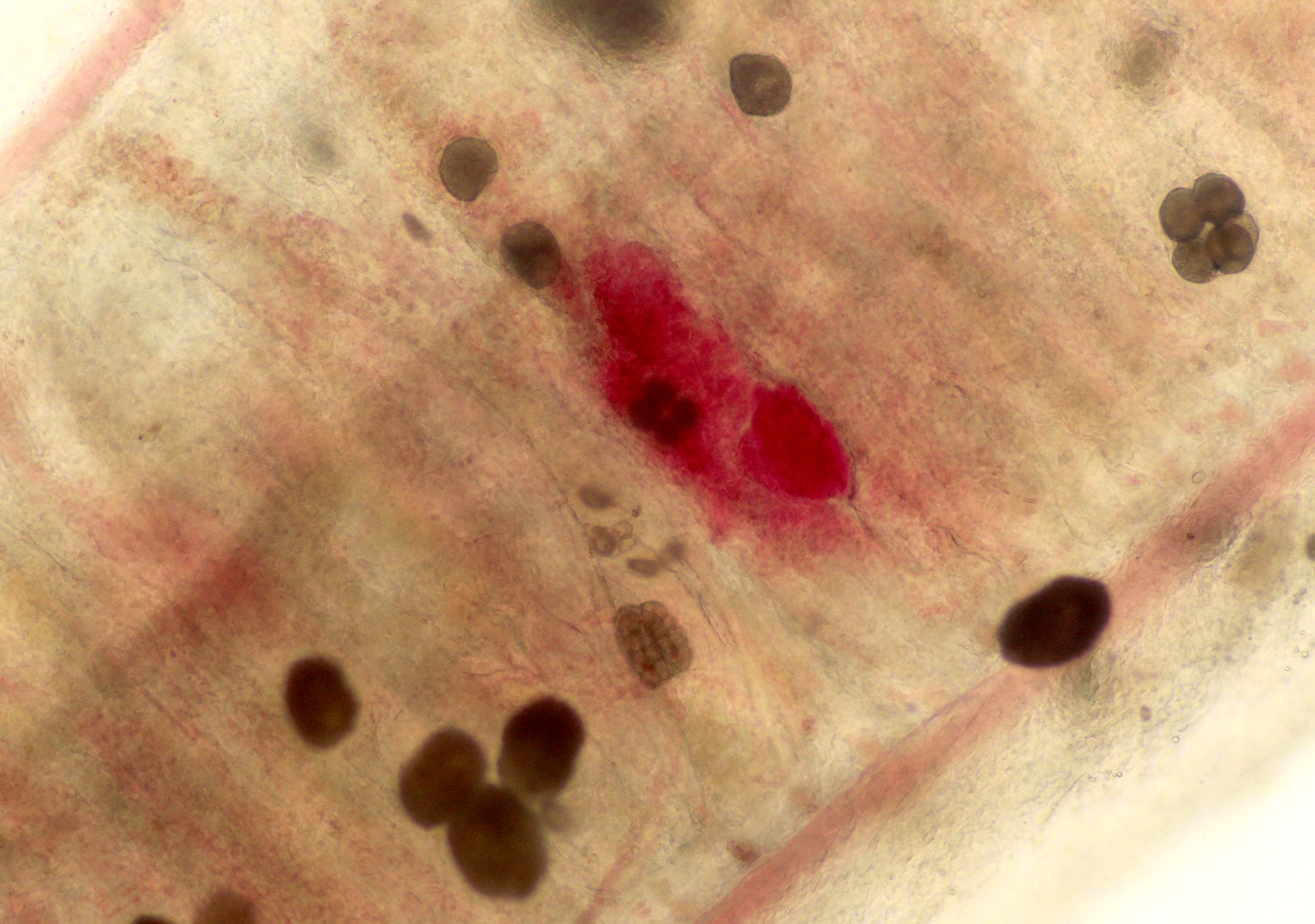
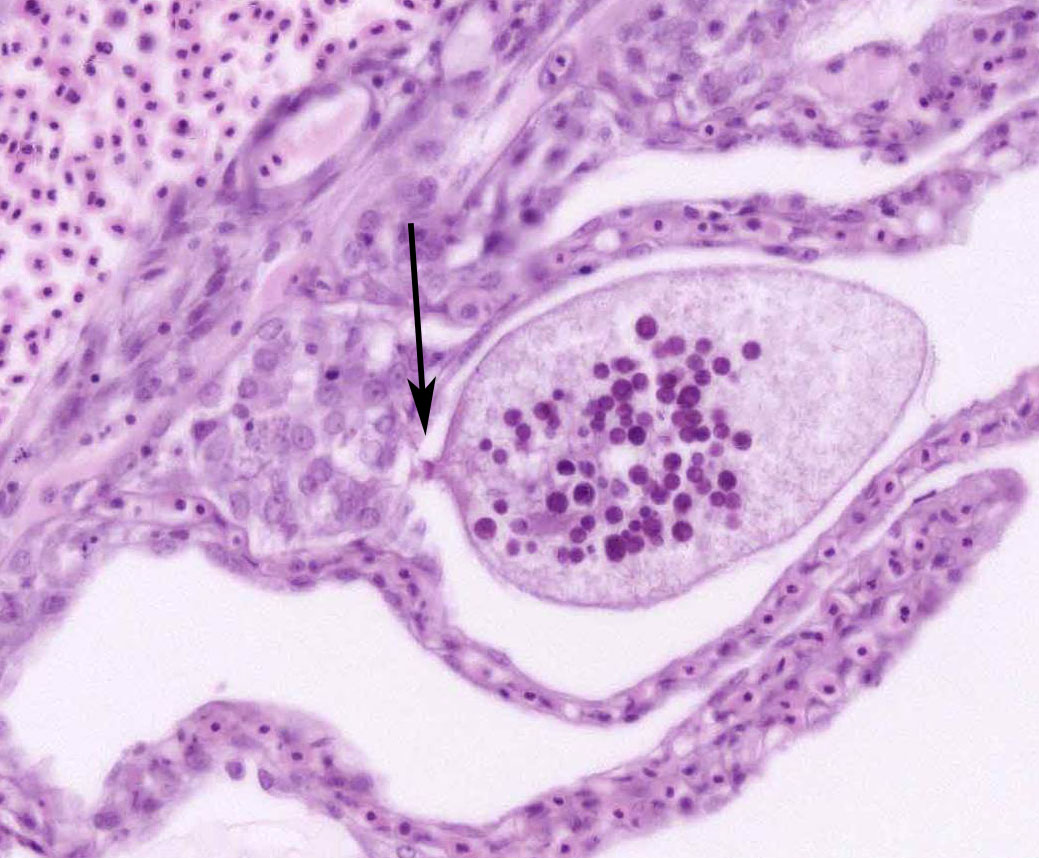
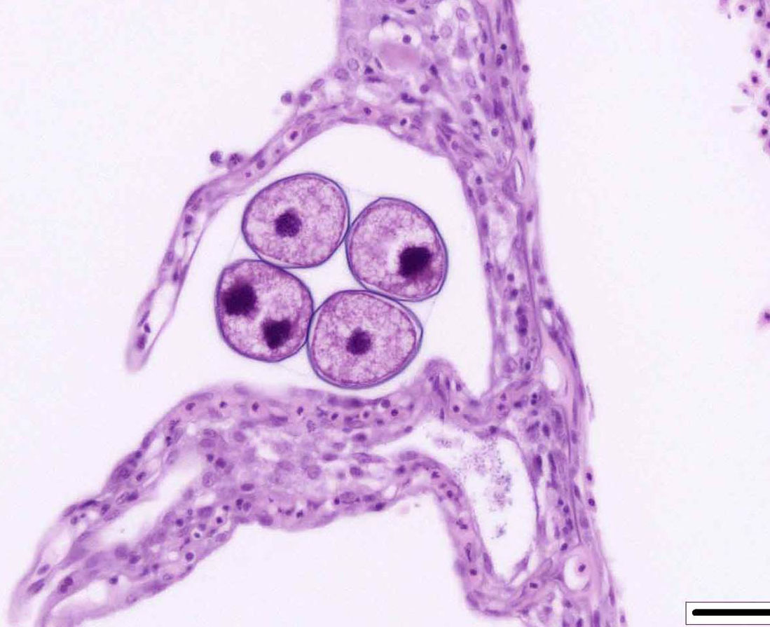
The life cycle of A. ocellatum is direct and triphasic. The parasitic, feeding stage, or trophont, attaches to the host�s epithelium by a root-like structured called a rhizoid.5 After a period of feeding on the host, the trophont detaches from the host, retracts its rhizoid and becomes the encysted, dividing tomont stage in the substrate.5 The final stage is the free-swimming, infectious dinospore that is released from the dividing tomont (up to 256 dinospores can come from a single tomont) and has flagella to facilitate swimming to find a suitable host.5 Amyloodinium ocellatum causes severe physical damage to the host cells through attachment of the trophont to epithelium, with the gill typically being the primary site of infestation.5,6 In cases with heavy parasite burdens, the skin and eyes can also be infected, sometimes producing the dusty appearance of the skin that accounts for the name �velvet disease.�5 In some reports infestation of larval fish affected only the skin rather than the gill.6 The gill was the tissue most severely affected in this puffer fish, although a few of the organisms were seen on a wet mount of the fin; cutaneous changes were mild in histologic sections. Trophonts may also be seen in the pseudobranch, branchial cavity, nasal passages, and gastrointestinal tract (if swallowed).5

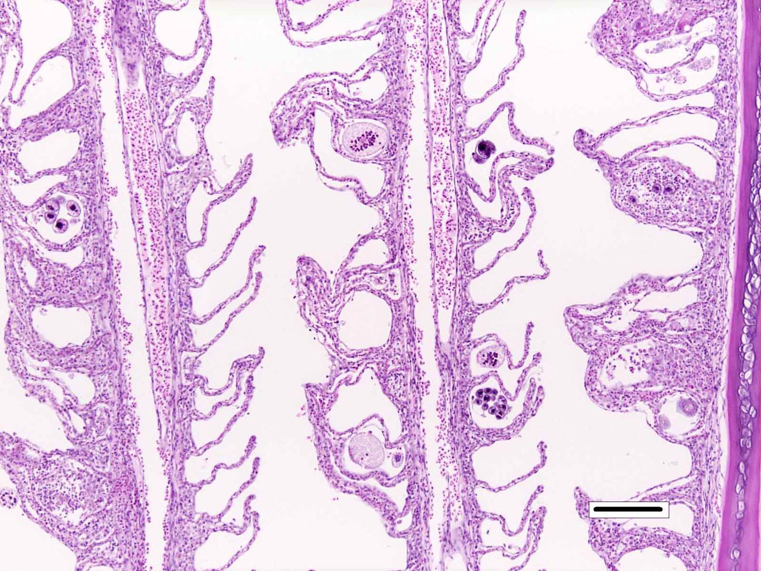
Microscopic lesions of severely affected gill include epithelial hyperplasia, often with lamellar fusion and distortion, mild inflammatory infiltrates, hemorrhage and epithelial degeneration and necrosis,3,5,6 as seen in this case. The feeding activity and the detachment of large numbers of organisms can damage epithelium, and potentially result in mortality as a consequence of hypoxia, osmoregulatory imbalance and secondary bacterial infections.5 There are several modalities of treatment available, most of which only target the dinospore stage, which can make eradication of parasites difficult.5

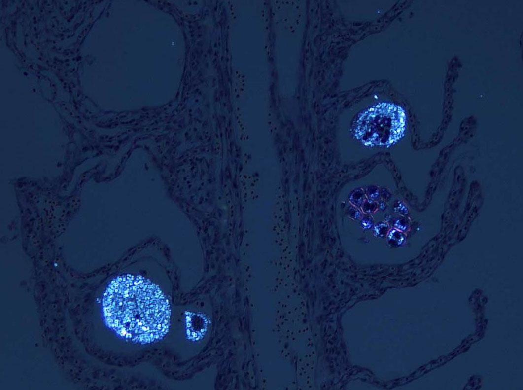
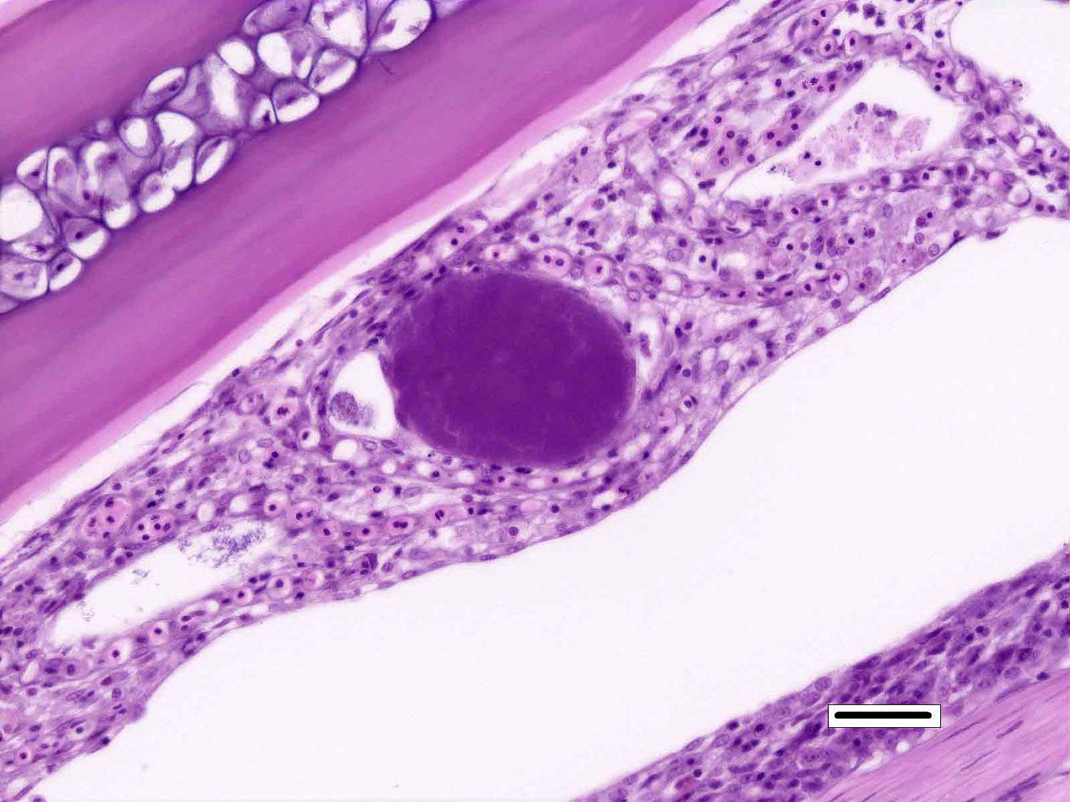
Although not considered the primary disease in this case, epitheliocystis was also noted in this fish. Characterized histologically by large, granular, basophilic, intracytoplasmic, bacterial inclusions within enlarged branchial lamellar epithelial cells, this type of infection is caused by a group of obligate, intracellular, gram-negative, chlamydia-like organisms.5 Recent advances in molecular techniques have identified various epitheliocystis agents as belonging to the order Chlamydiales, the first of which was Candidatus Piscichlamydia salmonis in the Atlantic salmon.1,7 Infections of gills, pseudobranch, and rarely the skin by these organisms have been described in many freshwater and marine species of fish.7 Pathogenicity is dependent upon the host response and the chlamydiales bacterium, and microscopic lesions can range from none to severe epithelial hyperplasia with associated inflammation.7


JPC Diagnosis:  

1. Gill: Branchitis, proliferative and necrotizing, multifocal, moderate with marked lamellar adhesions (synechiae) and fusion, and numerous dinoflagellate ectoparasites, Northern puffer fish, Sphoeroides maculatus.

2. Gill, lamellar epithelial cells: Chlamydial inclusions, multiple.


Conference Comment:  

Amylodinuym ocellatum is one of the most important pathogenic parasites of both marine and estuarine fish. It is one of the few organisms that can infect the gills and skin of both teleost fish (bony ray-finned fish) and elasmobranchs (cartilaginous fish, ex. sharks).2,5 This dinoflagellate parasite has the capacity for rapid reproduction and severe infections, such as in this case, can cause devastating disease and high mortality in a wide range of fish species.2 As nicely summarized by the contributor, the life cycle is simple and requires no intermediate hosts. The parasite has three different developmental stages. The trophont stage is the adult stage that feeds directly on the host and attaches via the rhizoid; the tomont stage is detached from the fish and divides to the third dinospore stage. Dinospores are the free swimming infectious flagellated phase.2,5 The simple life cycle and free swimming infectious dinospore stage contribute to severe epizootics in both wild and cultured fish, especially when crowded. A. ocellatum is a major concern to the aquaculture industry because outbreaks have an acute onset, spread rapidly, and have a high mortality. There are many nice examples of all three life stages in this case, including several trophonts with prominent rhizoids.2,5 This case is an excellent example of the gill as the primary site of infection for this parasite. Predominant antemortem clinical signs will most often be disturbances in the respiratory system, including increased respiratory rate (indicated by rapid movement of the gill opercula) and gathering at the surface or in other areas of increased oxygen concentration. The skin is also infected and fish can develop white or brown �velvety� appearance.2 This case also contains excellent examples of epitheliocystis with large inclusions composed of colonies of Chlamydia-like organisms within the cytoplasm of lamellar epithelial cells. Epitheliocystis is often considered an incidental finding, and most conference participants agreed that its presence in this case is unrelated to the significant gill pathology caused by Amylodinuym ocellatum.7

Prior to the conference, the moderator briefly reviewed the normal anatomy and histology of the gill. The gill arch is a series of bony or cartilaginous curved structures that support double rows of paired filaments, also called the primary lamellae. Each filament is composed of numerous perpendicularly-arranged secondary lamellae.4 The gill arch is covered by epidermis and at the origin of the primary lamellae the epidermis is thicker and contains many mucous cells and subjacent lymphoid tissue. The primary lamellae are covered by a mucoid epidermis which contains a pale-staining, eosinophilic, salt-secreting chloride cells that function in ionic transport and detoxification. The surface of the secondary lamellae consists of a single layer of interdigitating squamous epithelial cells supported by pillar cells and lamellar blood channels.4 The surface of the lamellar epithelium has numerous microvilli which serve as a substrate for cuticular mucus and aid in gas exchange and defense against infection and trauma. Thus, gills are important for gas exchange, ionic balance, and the excretion of the nitrogenous wastes. As a result, damage or infection of the gill often results in serious systemic con-sequences to the fish.4

Conference participants also discussed the difference between lamellar fusion and lamellar adhesion, both prominent features in this case. Lamellar fusion is most often associated with inflammation and is a consequence of marked epithelial proliferation. In contrast, lamellar adhesions (also known as lamellar clubbing or synechiae) occur when there is adherence of two or more secondary lamellae forming a pseudocyst, usually in the absence of epithelial proliferation or inflammation.  Both lamellar fusion and lamellar adhesions are present in this case.

Multifocally, within the secondary lamellae of this case, there are dilated capillaries that contain organizing fibrin thrombi, in-terpreted by conference participants as examples of telangiectasia. The moderator noted that this vascular change can be a perimortem artifact secondary to capture of the fish or an antemortem change secondary to the disease process. Distinguishing between peri- and antemortem telangiectasis is often difficult, even for those experienced in gill histopathology; however, in this case, the presence of organizing fibrin thrombi is characteristic of antemortem telangiectasia rather than a perimortem capture artifact.�


References:

1.      Draghi A, Popov VL, Kahl MM, et al. Characterization of �Candidatus Pischichlamydia salmonis� (Order Chlamydiales), a Chlamydia-like bacterium associated with epitheliocystis in farmed Atlantic Salmon (Salmo salar). J Clin Microbiol. 2004; 42(11):5286-5297.

2.      Francis-Floyd R, Floyd MR. Amyloodinium ocellatum, an important parasite of cultured marine fish. SRAC. 2011; 4705:1-12.

3.      Kuperman, BI, Matey VE. Massive infestation by Amyloodinium ocellatum (Dinoflagellida) in a highly saline lake, Salton Sea, California, USA. Dis Aquat Org. 1999; 39:65-73.

4.      Mumford S, Heidel J, Smith C, Morrison J, MacConnell B, Blazer V. Fish Histology and Histopathology. US Fish and Wildlife Service, National Conservation Training Center. http://nctc.fws.gov/resources/course-resources/fish-histology/index.html. Accessed May 25, 2017. 

5.      Noga EJ. Problem 27 Marine Velvet Disease (Amyloodiniosis, Marine Oodinium Disease, Oodiniosis). IN Fish Disease: Diagnosis and Treatment, 2nd ed, Ames, IA: Wiley-Blackwell; 2010:143-147.

6.      Paperna I, Steinitz AH. Amyloodinium ocellatum (Brown, 1931) (Dinoflagellida) infestations in cultured marine fish at Eilat, Red Sea: epizootiology and pathology. J Fish Dis. 1980; 3:363-372.

7.      Stride MC, Polkinghorne A, Nowak BF. Chlamydial infections of fish: Diverse pathogens and emerging causes of disease in aquaculture species. Vet Microbiol. 2014; 171:258�266.


Click the slide to view.


2-1. Gill, pufferfish.


2-2. Gill, pufferfish.


2-3. Gill, pufferfish.


2-4. Gill, pufferfish.


2-5. Gill, pufferfish.


2-6. Gill, pufferfish.


Back | VP Home | Contact Us |