Signalment:
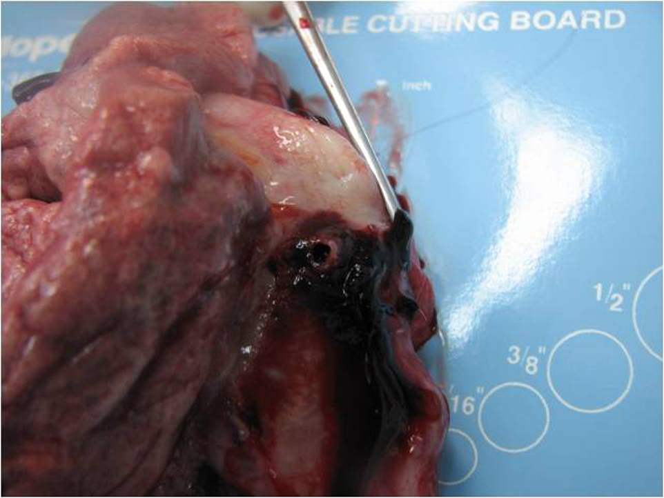
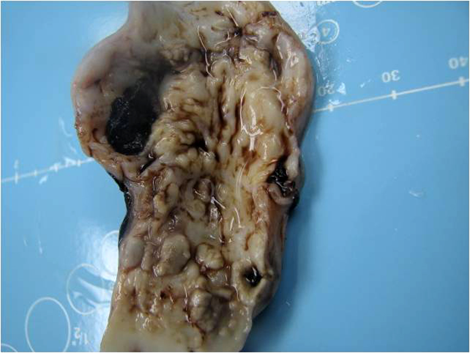
Gross Description:
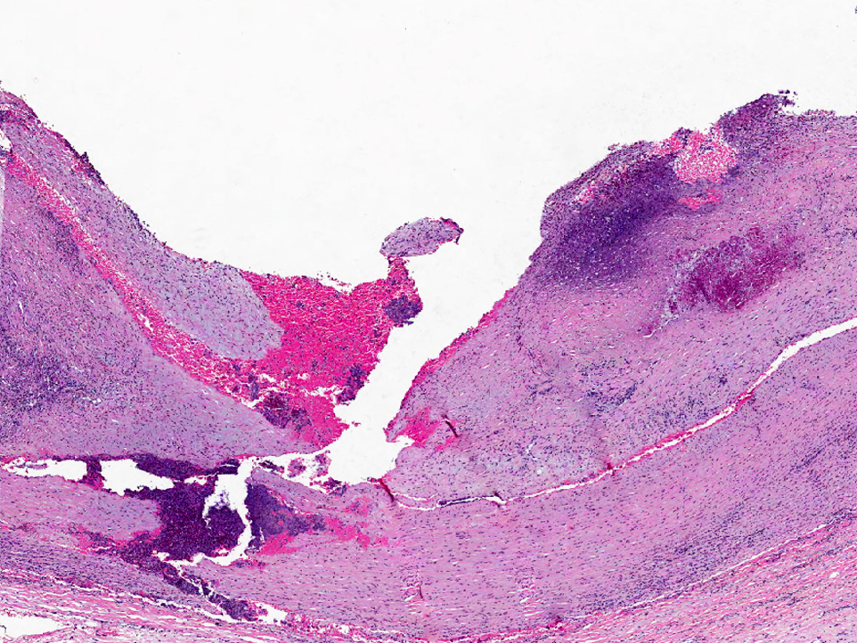
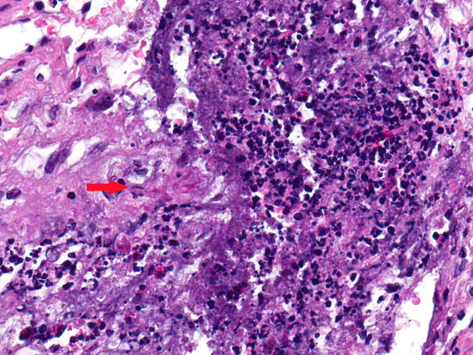
Histopathologic Description:
Morphologic Diagnosis:
Lab Results:
Condition:
Contributor Comment:
Aneurysm in the aortic arch associated with Aspergillus infection has been reported in a horse and fungal infections of the guttural pouch have been associated with ulceration of the internal carotid or maxillary artery resulting in their rupture and fatal hemorrhage in horses(2,7). Though spontaneous aortic aneurysms have been described in NHPs along with dissecting aneurysms in monkeys used as experimental models of atherosclerosis(6), our case uniquely presents rupture of a spontaneous aortic aneurysm associated with entomophthoromycosis in a sooty mangabey. Immunohistochemistry detected a mucormycete infection in the aorta and the PCR further identified the fungus as Basidiobolus spp, a mucormycete.Â
Broadly, the term mucormycosis is the preferred name used to describe the angiotropic infection caused by a member of the subphylum Mucoromycotina or the subphylum Entomophthoromycotina (formerly Zygomycetes). Histopathology typically demonstrates angioinvasion with associated necrosis(1). Medically important orders and genera include:
1. Mucorales, causing subcutaneous and systemic zygomycosis (Mucormycosis) - Rhizopus, Lichtheimia (Absidia), Rhizomucor, Mucor, Cunninghamella, Saksenaea, Apophysomyces, Cokeromyces and Mortierella.
2. Entomophthorales, causing subcutaneous zygomycosis (Entomophthoromycosis) - Conidiobolus and Basidiobolus(1)
Basidiobolus is a true pathogen, causing infections in immunocompetent and immunocompromised human and animal hosts(3). It occurs in decaying vegetation, soil, and as a saprobe in the intestinal contents of reptiles like lizards, chameleon, amphibians (toads) and mammals (bat)(3). The fungus is believed to enter the skin after insect bites, scratches and minor cuts. With the exception of Basidiobolus ranarum, the fungi in the Entomophthorales order, compared to those in Mucorales order, generally do not invade vascular tissue(3). Tissue reaction to Basidiobolus infection may be acute with infiltrates of eosinophils, lymphocytes and plasma cells at the infected focus and/or a chronic granulomatous response with a predominance of eosinophils.
Finally, the aneurysm in the thoracic aorta of this sooty mangabey was a true aneurysm involving all the layers of the aortic wall and in the absence of any significant clinical signs and based on the gross and histologic lesions, the cause of death of this animal was an acute rupture of the aortic aneurysm associated with the fungal infection. The mucormycotic infection of the aorta was very severe and multifocal, but no evidence of a widespread infection of other organs was observed.
JPC Diagnosis:
Conference Comment:
Zygomycetes are ubiquitous, saprophytic, non-pigmented, 7-10um wide fungi with non-dichotomous branching and rare septa. The Mucorales order is more commonly pathogenic in warm-blooded animals, with angioinvasion being characteristic, while the Entomophthorales order, which usually infects arthropods, reptiles, and amphibians, is rarely pathogenic to warm-blooded animals. Recent taxonomic restructuring combines the previous four species of the genus Basidiobolus into the single species, B. ranarum. The typical mode of transmition of Basidiobolus and other Entomophthorales is via ingestion of insects, which carry the spores on their external bristles, by reptiles and amphibians, which then pass infective spores in their excreta(11).
Differential diagnoses for these fungal hyphae should include Pythium and Lagenidium, members of the Oomycetes class(11). Polymerase chain reaction is needed to definitively differentiate these three pathogens. Another differential diagnosis that should be considered is the proliferative arteriopathy associated with SIV infection. With SIV, large to medium vessels are typically affected, particularly pulmonary arteries. The intima and media are thickened, the internal elastic lamina is fragmented, and he endothelium can be hypertrophied or hyperplastic, or focally eroded with adherent thrombi. Eosinophilic and granulomatous inflammation, as seen in this case, is typically not seen(5).
Some conference participants reported seeing Warthin-Finkeldey-type syncytial cells within germinal centers of the associated hyperplastic lymph node, which are occasionally seen in non-human primates infected with simian immunodeficiency virus (SIV) or measles virus, and represent the fusion of infected and uninfected CD4+ T lymphocytes. These cells have also seen in humans infected with HIV(9). Attempts to characterize these syncitial cells through immunohistochemistry and electron microscopy have been unfruitful, but they may represent a multinucleated follicular dendritic cell(10). While the presence of multinucleated cells was confirmed in the lymph node, the contributor confirmed that this sooty mangabey was seronegative for SIV.
References:
2. Ginn PE, Mansell, J.E.K.L., and Rakich, P.M. Skin and appendages, in Jubb, Kennedy, and Palmer's Pathology of Domestic Animals, M.G. Maxie, Editor. Elsevier Limited:Philadelphia, PA. p.707-608, 2007.
3. Gugnani HC. A review of zygomycosis due to Basidiobolus ranarum. Eur J Epidemiol 15:923-929,1999.
4. Jones TC, Hunt RD, King NW. Cardiovascular System. In: Jones TC, Hunt RD, King NW eds., Veterinary Pathology. 6th ed. Baltimore, MD: Williams & Wilkins; 1997:1001.
5. King NW. Simian Immunodeficiency Virus Infections. In: Jones TC, Mohr U, Hunt RD eds., Nonhuman Primates I. Berlin, Germany:Springer-Verlag; 1993:5-18.
6. Lowenstine, L.J., A primer of primate pathology: lesions and nonlesions. Toxicol Pathol, 31 Suppl: p. 92-102, 2003.
7. Okamoto M, Kamitani M, Tunoda N, Tagami M, Nagamine N, Kawata K, Itoh H, Kawasako K, Komine M, Akihara Y, Shimoyama Y, Miyasho T, Hirayama K, Kikuchi N, Taniyama H. Mycotic aneurysm in the aortic arch of a horse associated with invasive aspergillosis. Vet Rec 160:268-270, 2007.Â
8. Robinson, M.G.M.a.W.F., Cardiovascular System, in Jubb, Kennedy and Palmers Pathology of Domestic Animals, M.G. Maxie, Editor. Elsevier Limited: Philadelphia, PA. p. 62-63, 2007.
9. Rosenberg ES, Daskalakis DC. Impairment of HIV-specific immune effector cell function. In: ed. Badley AD. Cell death during HIV infection. Boca Raton, FL:CRC Press; 2006:195.
10. Sasseville et al. Induction of lymphocyte proliferation and severe gastrointestinal disease in macaques by a Nef gene variant of SIVmac238. AJP 1996; 149(1):163-176.
11. Songer JG, Post KW. Veterinary Microbiology, Bacterial and Fungal Agents of Animal Disease. St. Louis, MO:Elsevier Saunders; 2005:398-403.