WSC 2023-2024, Conference 17, Case 4
Signalment:
5-month-old, intact male Siberian Husky (Canis lupis familiaris)
History:
The dog had a history of progressive tetraparesis. Magnetic resonance imaging demonstrated bilaterally symmetric white matter hyperintensities in the corpus callosum, cerebrum, and thalamus. An analysis of the cerebrospinal fluid was normal. Given the breed and signalment, GM1 gangliosidosis was suspected and the dog was euthanized.
Gross Pathology:
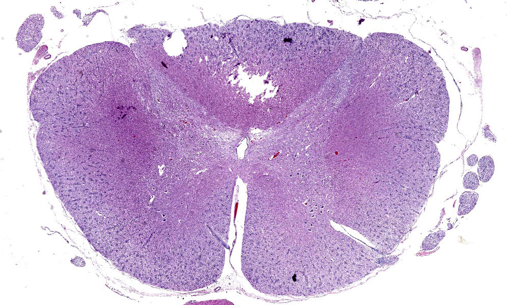
There was subtle flattening of the cervical spinal cord.
Laboratory Results:
DNA testing did not identify the gene mutation associated with GM1 gangliosidosis in Huskies.
Microscopic Description:
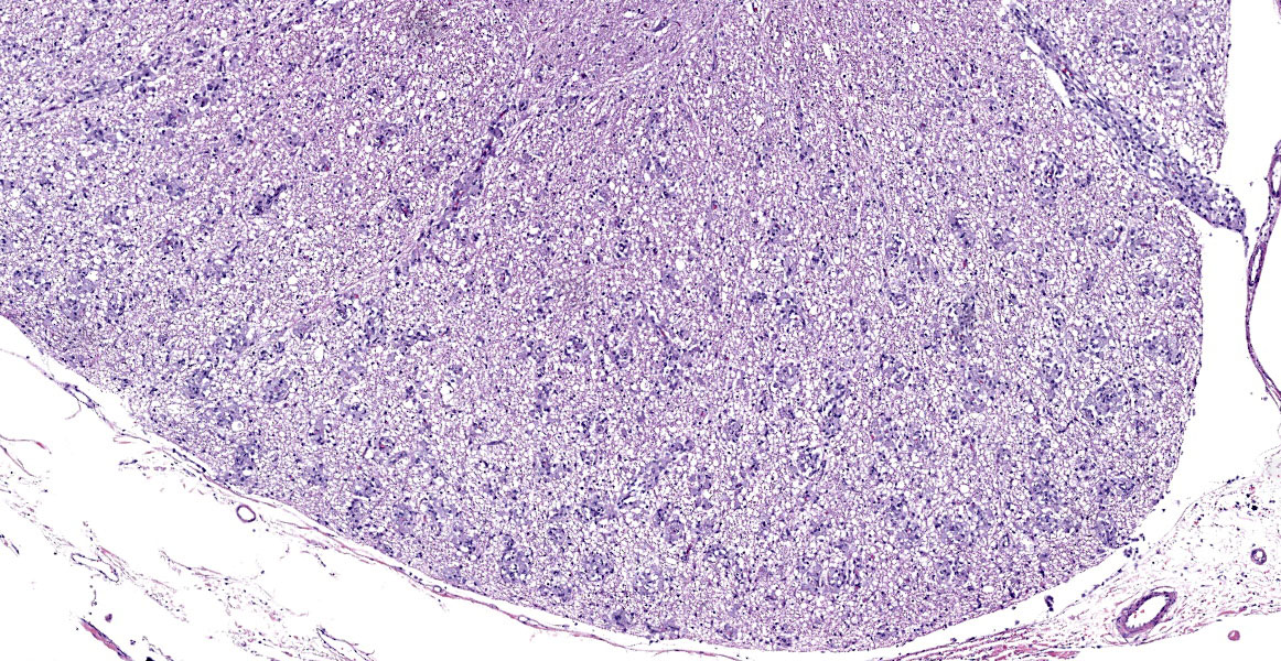
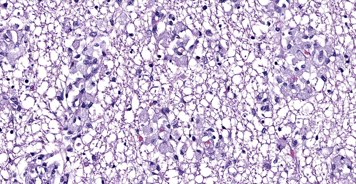
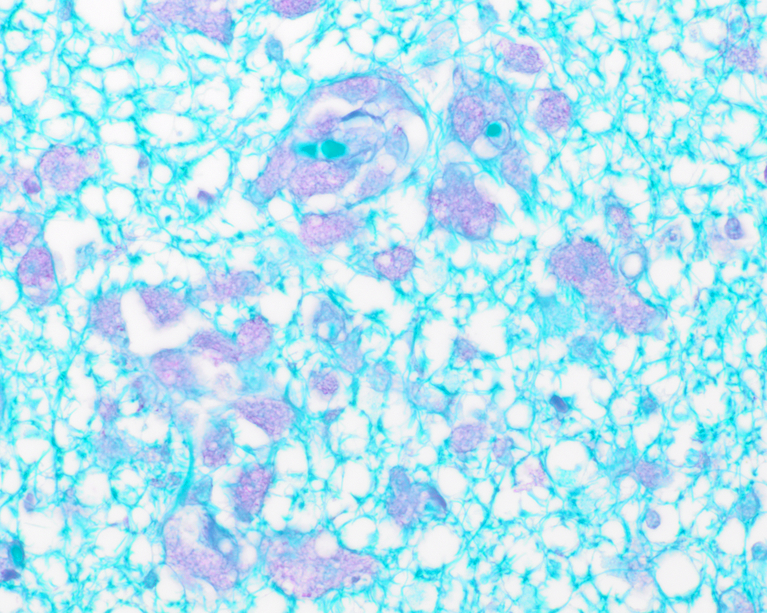
Cervical spinal cord: Within the white matter are aggregates of large round to polygonal cells with finely granular, lightly basophilic cytoplasm and eccentric nuclei (globoid cells). These cells occasionally surround blood vessels. The globoid cells contain faintly magenta material with a periodic acid- Schiff stain. There is marked loss of myelin and axons highlighted by Luxol fast blue and Sevier-Munger stains, respectively. The white matter in other portions of the brain and spinal cord were similarly affected.
Contributor’s Morphologic Diagnosis:
Marked widespread axonal degeneration with marked histiocytosis.
Contributor’s Comment:
The clinical signs, gross, and microscopic findings are attributed to axonal degeneration in the brain and spinal cord as a result of globoid cell leukodystrophy. Globoid cell leukodystrophy occurs due to deficient activity of the lysosomal enzyme galactocerebrosidase. Globoid cell leukodystrophy is a genetic condition reported in many pure breed dogs (Cairn Terriers, West Highland White Terriers, Miniature Poodles, Bluetick Hounds, Basset Hounds, Beagles, Irish Setters and the Australian Kelpie).1 It has not been reported in Siberian Huskies. Additionally, sheep, cats and primates may be affected.3
The pathophysiology of globoid cell leukodystrophy is complicated and not fully understood. Galactocerebrosidase normally breaks down galactocerebroside and galactosylsphingosine (psychosine).1 Interestingly, at the time of diagnosis, many individuals have decreased amounts of galactocerebroside; this paradox may reflect the decreased amounts of myelin remaining late in the disease course.2 There is, however, accumulation of psychosine in the oligodendroglia and Schwann cells. The psychosine is extremely toxic to these cells and results in their death. The loss of oligodendroglial cells and Schwann cells results in decreased myelination of axons and ultimately, axonal degeneration. Macrophages attempt to clear remaining myelin and galactocerebrosides, but without the enzyme they are also unable to break down galactocerebrosides. As a result, the macrophages become swollen with PAS-positive debris and are termed “globoid cells.”1 The globoid cells often accumulate around blood vessels, as in this case. Although a biopsy of peripheral nerves can be useful to make an antemortem diagnosis, in this case, the sciatic nerve was not clearly affected.1
Contributing Institution:
University of Tennessee
College of Veterinary Medicine
Department of Biomedical and Diagnostic Sciences
http://www.vet.utk.edu/departments/path/index.php
JPC Diagnosis:
Spinal cord, white matter: Histiocytosis, perivascular, diffuse, marked, with abundant intracellular myelin.
JPC Comment:
Globoid cell leukodystrophy (GLD), also known as Krabbe disease, is an autosomal recessive, fatal lysosomal storage disease caused by mutations in galactoceramide beta-galactosidase (also known as galactosylceramidase, galactocerebrosidase, or GALC). GLD develops early in life and, as noted by the contributor, is characterized by impaired aberrant myelin metabolism and the subsequent accumulation of galactosylceramide (GalCer) and psychosine, the major substrates of GALC.3
Compared with normal cellular membranes, myelin has a very high lipid content and is enriched in GalCer.2 Myelin is remarkably stable, but is contininously remodeled at a basal rate and at a faster rate in response to injury or disease. The complex myelin degradation process relies on a number of enzymes, including GALC, to break down its consitutent parts, including GalCer.2 GALC is expressed ubiquitously in the lysosomes of all cell types in the nervous system, and is particularly enriched in oligodendrocytes.2 In health, once GalCer is processed in the lysosome, its consitutent molecular parts are recycled and become building blocks in the constitutively active myelin remodeling/remyelination pathway.3
In GLD, mutations diminish GALC enzyme activity, resulting in impaired degradation of GalCer during myelin remodeling. The resulting lack of sufficient building blocks for remyelination leads to myelin loss.3 Concurrently, toxic pysochosine, normally produced by oligodendrocytes and also degraded by GALC, begins to accumulate, leading to extensive oligodendrocyte degeneration and death with consequent degeneration of existing myelin and impairment of remyelination.1
As the contributor notes, macrophages arrive to clean up the degenerating myelin, but they are also GALC-deficient and cannot metabolize GalCer. The acc
The globoid cells appear in perivascular cuffs in the white matter of the CNS, in the leptomeninges, and in the endoneurium of peripheral nerves.1
This case is an excellent example of the pathognomonic histology of GLD, characterized by marked myelin loss, markedly reduced numbers of oligodendrocytes, and, most dramatically, the infiltration of numerous large, round macrophages filled with PAS-positive storage material, most abundant in areas of active demyelination.3 The presenting clinical signs in this case–ataxia and limb weakness and tremors that progress to paralysis and muscular atrophy–are also typical.4 Some animals may also develop blindness. Typical gross lesions include slight gray discolorations of the white matter, particularly in the centrum semiovale of the cerebrum and in the spinal cord.4
Conference participants enjoyed the photogenic and straightforward nature of this case. Dr. Miller cautioned residents not to overinterpte the gray matter vacuolation present in some sections of the spinal cord since, as in so many neuroparenchymal lesions, this likely represents autolysis artifact. Dr. Miller discussed leukodystrophies in general, noting that the various diseases have in common a failure of myelinating cells to maintain their myelin sheaths. Such diseases are typically early onset, symmetrical, and typically unaccompanied by significant inflammation.
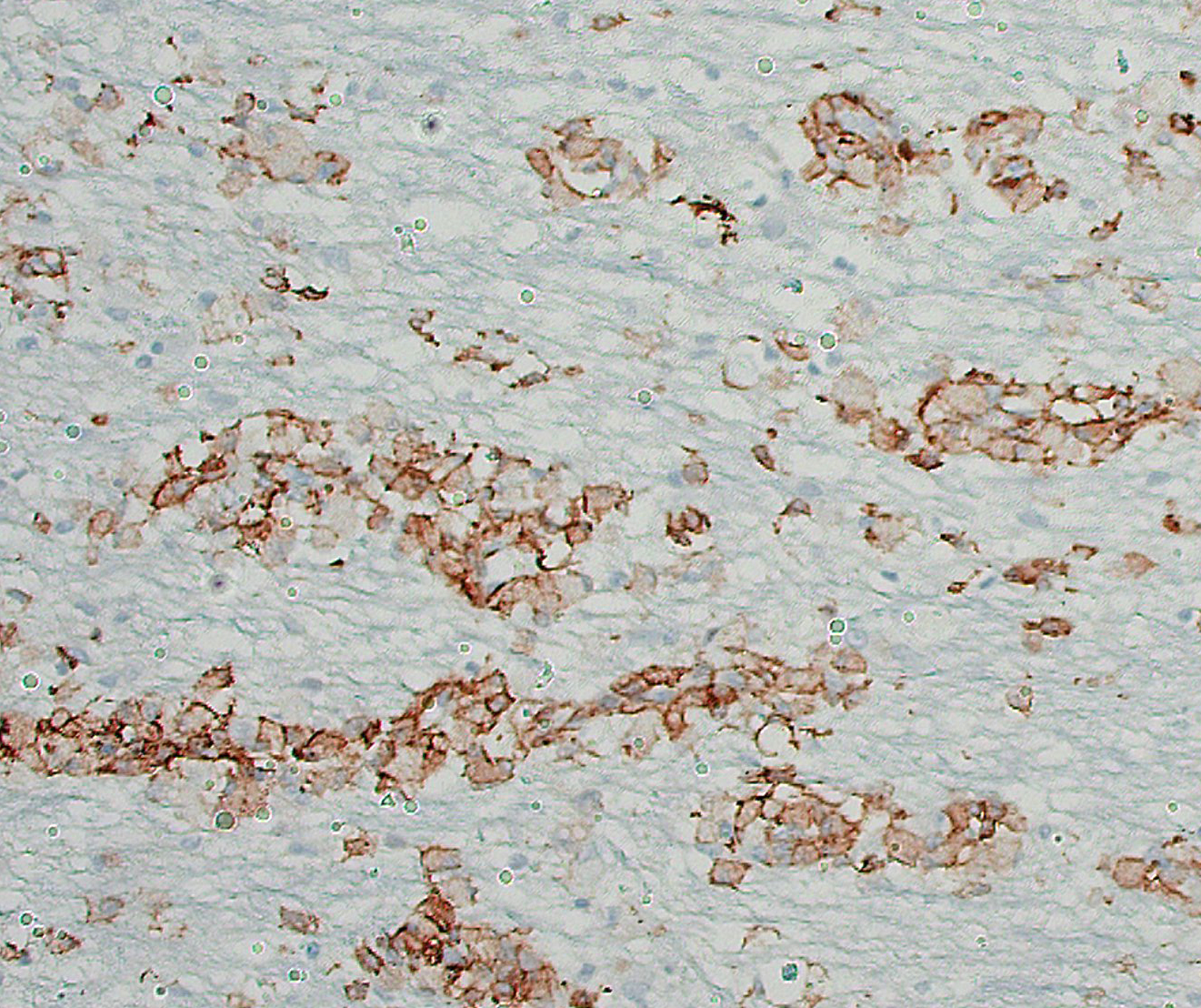
Dr. Miller briefly reviewed the biology and function of the lysosome as well as sphingolipid metabolism and the diseases that result from associated enzyme deficiencies. Dr. Miller discussed the histochemical staining characteristics of globoid cells and noted that IBA1, an immunohistochemical stain for macrophages; PAS, which stains the material accumulated in globoid cells; and Olig2, an immunohistochemical stain for oligodendrocytes, can be used in concert to demonstrate that the accumulation in GLD is occurring in within histiocytes and not oligodendrocytes.
References:
- Cantile C and Youssef S. Chapter 4: Nervous System. In: Maxie MG ed. Jubb, Kennedy and Palmer’s Pathology of Domestic Animals. 6th ed. vol. 1. Elsevier; 2016.
- Feltri ML, Weeinstock NI, Favret J, Dhimal N, Wrabetz L, Shin D. Mechanisms of demyelination and neurodegeneration in globoid cell leukodystrophy. Glia. 2021. 69:2309-2331.
- Lee E, Fuller M, Carr M, Manavis J, Finnie J. Globoid cell leukodystrophy (Krabbe’s disease) in a Merino sheep. J Vet Diagn Invest. 2019;31:118-121.
- Miller AD, Porter BF. Nervous System. In: Zachary JF, ed. Pathologic Basis of Veterinary Disease. 7th ed. Elsevier; 2022:945-946.