Joint Pathology Center
Veterinary Pathology Services
Wednesday Slide Conference
2019-2020
Conference 2
28 August 2019
CASE II: 16GR108 (JPC 4118174).
Signalment: 1-year-old, male, Beagle (Canis familiaris).
History: Two-week exploratory toxicity study.
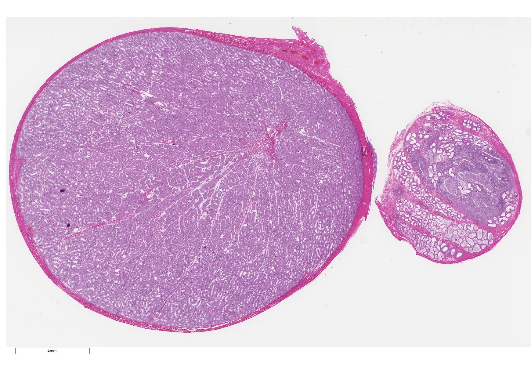
Gross Pathology: Unilateral (left) small epididymis was noted macroscopically.
Laboratory results: None
Microscopic Description:
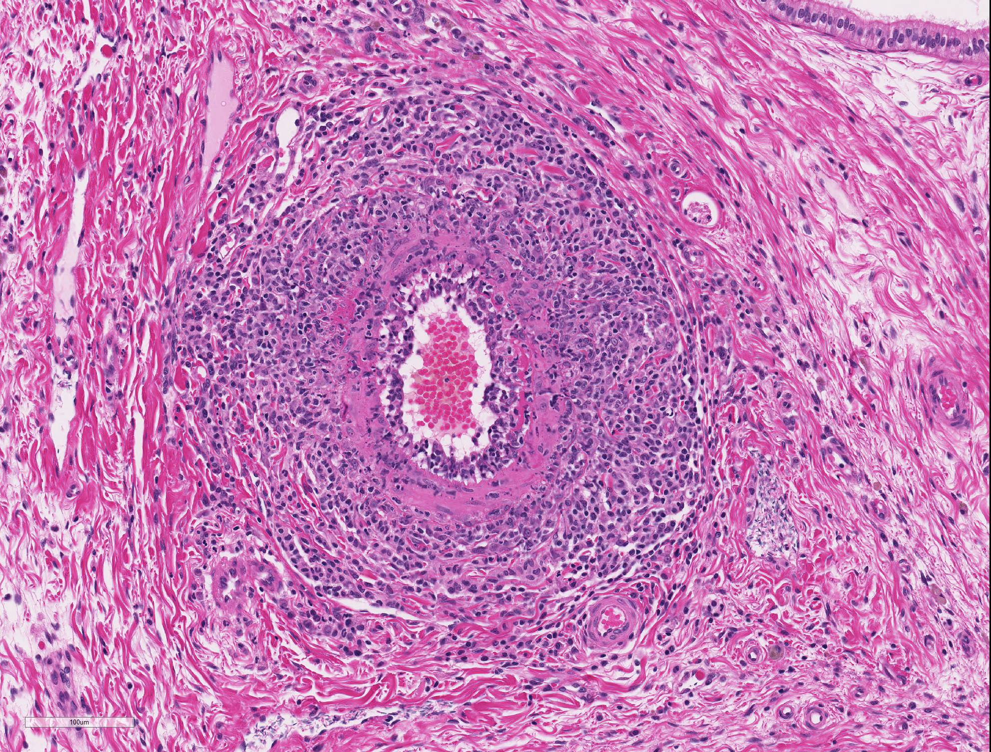
Epididymis: Within the epididymis, there is moderate focal arterial inflammation characterized by expansion of tunica intima with prominent endothelial cells, disruption of the internal elastic lamina, and presence of fibrin, cellular debris, and inflammatory cells (lymphocytes, plasma cells and macrophages) in the tunica media and adventitia.
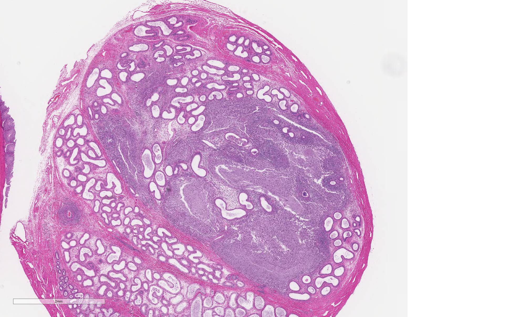
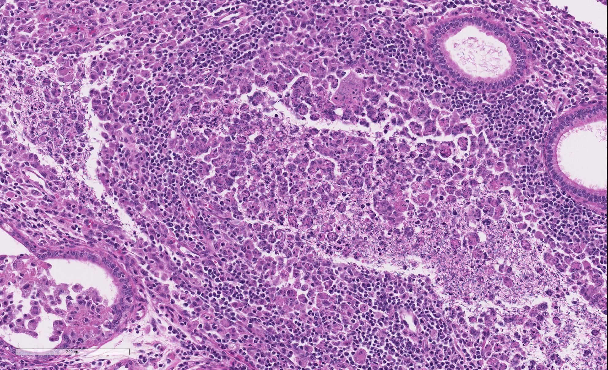
Disrupting ductular and stromal architecture is a focally extensive inflammatory reaction composed of a ring of macrophages, lymphocytes, and plasma cells (granuloma) surrounding a large focus of extravasated spermatozoa.
Contributor
Morphologic Diagnoses:
Epididymis: 1)
Arteritis, lymphoplasmacytic, focal, moderate, subacute
2) Sperm granuloma, focal, moderate, subacute
Contributor Comment:
This finding was unrelated to the test article because it occurred in just one animal without dose relationship and was consistent with idiopathic juvenile polyarteritis of young Beagles, a background finding in dogs.1,2 Small to medium caliber arteries are most commonly affected by this form of vascular injury. Histologically, vascular changes are characterized by medial necrosis, mononuclear cell inflammatory infiltrates, intimal proliferation, and fibrinoid necrosis.4,7,8 There is a higher incidence in males with a trend towards the epididymis as the organ most commonly affected. Differentiation from drug-induced vascular injury (DIVI) is important in toxicity studies.
DIVI includes vasoactive arteriopathy, toxic vasculitis, and hypersensitivity vasculitis. Histologic changes induced by vasoactive drugs include necrosis and inflammation in all three vessel layers, most commonly in small vessels of the skin, and an absence of fibrinoid necrosis. Toxic vasculitis affects small to medium-caliber vessels and is characterized morphologically by segmental fibrinoid necrosis and neutrophilic inflammation of the vessel wall, periadventitial tissue, and thrombosis. Vasoactive arteriopathy represents the most common type of DIVI and primarily affects coronary arteries (vasodilators) and small arteries systemically (vasoconstrictors). Histologically, changes are characterized by medial hemorrhagic necrosis with minimal inflammation (vasodilators) and focal medial fibrinoid necrosis (vasoconstrictors).1
Epididymal sperm granulomas are typically an incidental finding and similarly not test article-related in the present case. There are multiple causes of sperm granulomas including, most commonly, trauma and inflammation, congenital ductular abnormalities, adenomyosis of epididymal ducts, parasitic lesions, and toxins.2
Contributing Institution:
Pfizer Drug Safety Research and Development
455 Eastern Point Rd.,
Groton, CT 06340
https://www.pfizer.com/partners/research-and-development
JPC Diagnosis:
1.
Epididymis: Sperm granulomas, multiple.
2. Arteriole, head of the epididymis: Arteritis, necrotizing and
proliferative, diffuse, moderate.
3. Testis, seminiferous tubules: Atrophy and aspermatogenesis, diffuse,
moderate.
JPC Comment: Idiopathic juvenile polyarteritis of beagle dogs, first described in 1992 by Scott-Moncrieff et al. in 1992,6 is one of the most well-known primary vasculitides in the dog. The term "primary" vasculitis comes from human medicine and refers to a vasculitis whose cause is unknown, versus "secondary" vasculitides, which have defined infectious causes, such as endotheliotropic viruses, or drugs (as described by the contributors). A retrospective study of canine vasculitis by Swann et al. in 20159 failed to disclose significant differences between the two groups; primary vasculitides were more common in female dogs (this case notwithstanding), and dogs with elevated serum globulin concentrations were seen with more frequency in dogs with primary vasculitides.
Idiopathic
juvenile polyarteritis (IJP), also known as "beagle pain syndrome", is an acute
febrile multisystemic vasculitis most often seen in dogs less than 36 months of
age.7 It is characterized by acute onset of fever and pain (often
cervical) with animals assuming a characteristic hunched stance.4
Vascular lesions range from a mild lymphohistiocytic perivasculitis to
transmural arterial inflammation with fibrinoid necrosis with loss of elastic
laminae, intimal and medial fibrosis, and marked perivascular inflammation.4,7
Lesions are most commonly seen in the small-and medium-sized arterioles (i.e.,
arterioles other than the great vessels) of the cervical meninges, heart, and
cranial mediastinum.7 Animals with repeated episodes may develop
gross signs of progressive bilateral atrophy of cervical and temporal muscles,4
and histologic evidence of hepatic, splenic and renal amyloidosis.4,7
Interestingly, a primary vasculitis affecting the testicular artery of raccoons
has been reported.5 Histologic lesions of segmental arteritis of the
extratesticular (epididymis and spermatic cord) arterioles similar to that of IJP
and other forms of polyarteritis nodosa were described. The lesions were seen
in both wild and breeder animals of all ages.5
The blood-epididymis barrier (BEB) is an important factor in protecting maturing sperm from the immune system. This barrier protects developing spermatozoa in the testis and mature spermatozoa within the epididymal tubules not only from infiltrating inflammatory cells following injury, but also from cytokines released in local and systemic inflammatory conditions.3 The breakdown of the BEB may result from traumatic injuries, developmental abnormalities, or the administration of testosterone and various chemicals and drugs. In addition, BEB barrier function diminishes with age. Sperm granulomas (which may be seen not only in the epididymis but also within the testis proper and vas deferens) are cardinal signs of breakdown of the BEB, and arise as a result of the antigenicity of spermatozoa.3 An excellent review of factors contributing to the breakdown of the BEB and the resulting inflammatory process is provided in the 2014 article by Gregory and Cyr cited below.3
The moderator
discussed the formation of sperm granuloma and the fact that the inception of
spermatozoa formation does not occur until well after the immune system is
formed, which is why sperm is so antigenic. She pointed out that the tail of
the epididymis is more likely to be affected in infectious disease, while the
head (caput) of the epididymis has a higher incidence of granuloma formation as
a result of the presence of an increased number of abnormal ductules and
reviewed the general pathology behind granuloma formation. She gave an
excellent review of "beagle pain" syndrome, pointing out that the disease is
not restricted to Beagles, nor is it always a painful condition. She chose
this case as an example of a classic lesion as well as a descriptive case which
has two concurrent processes.
References:
1. Clemo FAS, Evering WE, Snyder PW, et al. Differentiating spontaneous from drug-induced vascular injury in the dog. Toxicol Pathol. 2003;31(Suppl.):25-31.
2. Foley GL, Bassily N and Hess RA. Intratubular spermatic granulomas of the canine efferent ductules. Toxicol Pathol. 1995;23(6): 731-734.
3. Gregory M, Cyr DG. The blood-epididymis barrier and inflammation. Spermatogenesis 2014; 4(2)e97619-1 â e97619-13.
4. Hayes TJ, Roberts GK, Halliwell WH. An idiopathic febrile necrotizing arteritis syndrome in the dog: beagle pain syndrome. Toxicol Pathol. 1989;17(2):129-137.
5. Hamir AN, Palmer M, Li H, Stanko J, Rogers DB. Spontaneous idiopathic arteritis of the testicular artery in raccoons (Procyon lotor). Vet Pathol 2009; 46:1129-1132.
6. Scott-Moncrieff JC, Snyder PW, Glickman LT, Davis EL, Felsburg PJ. Systemic necrotizing vasculitis in nine young beagles. J Am Vet Med Assoc 1992; 201(10):1553-1558.
7. Snyder PW, Kazacos EA, Scott-Moncrieff JC, et al. Pathologic features of naturally occurring juvenile polyarteritis in beagle dogs. Vet Pathol 1995;32(4):337-345.
8. Son W. Idiopathic canine polyarteritis in control beagle dogs from toxicity studies. J Vet Sci. 2004;5(2):147-150.
9. Swann JW, Priestnall SL, Dawson C, Chang YM, Garden OA. Histologic and clinical features of primary and secondary vasculitis: a retrospective study of 42 dogs. J Vet Diagn Investig 2015; 27(4): 489-496.