Signalment:  

1 -+ years, female, rabbit (Oryctolagus cuniculus)The rabbit was presented with symptoms of salivation, seizures, somnolence and fever. Meningitis was suspected.


Gross Description:  

Yellowish covering of the eye-¦s proximity; ulceration of the right cornea; edema of the mucosal-cutaneous intersections; slight enlargement and blood-reabsorption of the lymph nodes at the head; little yellowish dry mass in the outer ear canals; no abnormalities at the inner ears; slight lipidosis of the liver; dilated uterus, filled with white mucous. 


Histopathologic Description:

Brain: multifocal moderate to severe perivascular accumulation of lymphocytes, plasma cells and histiocytes at meningeal and cortical blood vessels; multifocal extensive neuronal and glial necrosis at the cerebral cortex; detection of intranuclear eosinophilic to amphophilic inclusion bodies in cortical pyramidal neurons and glial cells, often filling the entire nucleus; electron microscopy showed high numbers of intranuclear particles of icosahedral nucleocapsids, consistent with the morphology of herpesvirus.

Eye (not on the slide): ulceration of the cornea; small foci of infiltrating lymphocytes and plasma cells at the limbus.


Morphologic Diagnosis:  

Brain: Meningoencephalitis, severe, multifocal, non-suppurative, with detection of numerous intranuclear inclusion bodies in neurons and glial cells. 


Lab Results:  

Herpes simplex virus (HSV) was verified by polymerase chain reaction of paraffin-embedded, formalin-fixed brain material. Specificity of HSV-1, was confirmed by restriction enzyme digestion with BamHl and Sma-digestion (Institute of virology, TU Munich, Germany).


Condition:  

Meningoencephalitis


Contributor Comment:  

Herpesvirus infection is an uncommon disease in domestic rabbits. Herpesvirus sylvilagus and cuniculi (formerly known as virus III or herpes-like virus) are known to be either only pathogenic for individual breeds or only slightly pathogenic for the domestic rabbit.(5,2) A third, unclassified herpesvirus causes haemorrhagic dermatitis, pneumonia and necrotizing splenitis in rabbits.(6) Naturally occurring encephalitis in domestic rabbits by herpes simplex virus infection has only been reported twice.(9,1) In both reported cases a person with herpetic infection and close contact to the rabbit was suspected to be the source of infection for the animal.

Rabbits can be easily infected experimentally and act as an animal model for human herpes simplex infection.(7) Trigeminally innervated areas, as for example the cornea, serve as the portal of entry for the human herpes simplex virus.(7) Furthermore, nasal infection is known to lead to focal lesions in the brain.(8)


JPC Diagnosis:  

Cerebrum: Neuronal necrosis, multifocal, with intranuclear inclusion bodies and mild lymphocytic meningitis.


Conference Comment:  

Rabbits serve as experimental models for herpes simplex virus (HSV) type 1 (human herpesvirus 1) encephalitis in humans, and in rabbits, the infection is exclusively neurotropic. In humans, the intranasal route of infection is most important in development of encephalitis. Experimental intranasal ino-culation in rabbits leads to migration via the olfactory nerves, leading into the frontal and temporal lobes. Intraocular inoculation of the virus in rabbits has also been used to study cell spread of HSV, and rabbits can present with neurologic signs as early as 2 days post infection; the disease is also known to progress quickly in spontaneous cases.(3) Following intraocular inoculation, the virus travels through the optic nerve to the corpus geniculatum.(1,3) Both ocular and nasal routes of inoculation can lead to seizures and death but the intranasal route has a higher mortality rate. Histologic lesions described in other cases of both natural and experimental infection are similar to those seen in this case.(1,3,9) Gross CNS lesions in rabbit HSV infections are uncommon which may be due in part to the rapid course of disease. In some reported cases of natural HSV infection in rabbits, humans in close contact were reported to have cold sores prior to the onset of clinical signs in the rabbit. Nonetheless, it is unclear if spontaneous HSV encephalitis in rabbits can arise from reactivation of latent infection or if disease only occurs shortly after exposure.(3)

HSV can also result in fatal disease in nonhuman primates. In Old World primates, the course of HSV infection is comparable to humans with localization to the mucocutaneous tissues and relatively mild disease. However, New World primates (NWP) are considered highly susceptible and infection often leads to severe systemic disease and death with a rapid clinical course in many cases. In some NWPs such as owl monkeys and marmosets, they also develop ulceration of the oral mucous membranes but it is accompanied by hemorrhage and necrosis in the cerebral cortex as well as many other organs. In most of the reported cases in non-human primates, close contact with an infected human was found to be the source of infection. The most characteristic lesions in HSV infection in NWPs are oral ulcerations and lesions at the mucocutaneous junction, which cannot be grossly differentiated from lesions caused by herpesvirus T (Herpes tamarinds) infection. Molecular methods are needed to differentiate infections caused by the two viruses due to similarity of lesions.(4)

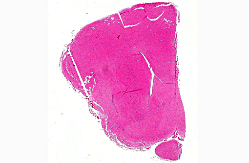
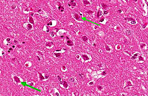
The conference description included multifocal, random areas of neuronal necrosis within the superficial gray matter accompanied by gliosis, satellitosis and low numbers of infiltrating heterophils. Perivascular cuffing by mononuclear cells is present multifocally within the gray matter and a similar mononuclear infiltrate as well as edema expand the meninges. The lack of neuronal degenerative changes such as swelling was noted by some participants; however, in some viral infections, neuronal necrosis (even in acute infection) may be the defining lesion.


References:

1. Grest P, Albicker P, Hoelzle L, Wild P, Pospischil A. Herpes simplex encephalitis in a domestic rabbit (Oryctolagus cuniculus). J Comp Pathol. 2002; 126: 308-11.

2. Hinze HC. New Member of the Herpesvirus Group Isolated from Wild Cottontail Rabbits. Infect Immun. 1971; 3: 350-354.

3. Nesburn AB. Isolation and characterization of a herpes-like virus from New Zealand albino rabbit kidney cell cultures: a probable reisolation of virus 3 of Rivers. J Virol. 1969; 3: 59-69.

4. Matos R, Russell D, Van Alstine W, Miller A. Spontaneous fatal Human herpesvirus 1 encephalitis in two domestic rabbits (Oryctolagus cuniculus). J Vet Diag Invest. 2014; 26(5):689-694.

5. M+â-ñtz-Rensing K, Jentsch KD, Rensing S, Langenhuyzen S, et al. Fatal Herpes simplex infection in a group of common marmosets (Callithrix jacchus). Vet Pathol. 2003; 40:405-411.

6. Onderka DK, Papp-Vid G and Perry AW: Fatal herpesvirus infection in commercial rabbits. Can Vet J. 1992; 33:539-543.

7. Paeivaerinta MA, Roeyttae M, Hukkanen V, Marttila RJ. Nervous system inflammatory lesions and viral nucleic acids in rabbits with herpes simplex virus encephalitis-induced rotational behaviour. Acta Neuropathol. 1994; 87: 259-68.

8.  Stroop WG and Schaefer DC. Production of encephalitis restricted to the temporal lobes by experimental reactivation of herpes simplex virus. J Infect Dis. 1986; 153: 721-731. 

9. Weissenboeck H, Hainfellner A, Berger J, Kasper I and Budka H. Naturally occurring herpes simplex encephalitis in a domestic rabbit (Oryctolagus cuniculus). Vet Pathol. 1997; 34: 44-7.


Click the slide to view.


3-1. Cerebrum


3-2. Cerebrum


3-3. Neuron


Back | VP Home | Contact Us |