Signalment:
20-year-old, gelding, Arabian horse (
Equus caballus)A cecal mass was submitted for histopathology.
Gross Description:
The submitted tissue was an approximately 4x5x4 cm round, firm, nodular, dark gray, well encapsulated mass with intact surface mucosa. On cut section the mass was distinct from the mucosa, white-tan and multilobulated.
Histopathologic Description:
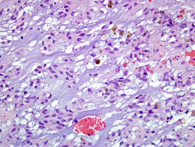
The mass is a well demarcated, encapsulated, multilobulated, expansile mass located from deep tunica muscularis to the serosa compressing the adjacent tissues and muscle layers. The mass is composed of multiple large lobules separated by a thick dense fibrous connective tissue. The mass is made of a large number of spindle shaped cells arranged in interconnected and branching trabeculae separated by sinuses filled with large amount of basophilic mucinous material as well as clusters of erythrocytes (
Fig. 1-1). The neoplastic cells have indistinct cell borders, large amount of eosinophilic cytoplasm, one oval to elongated nucleus with rounded poles and finely granular heterochromatin and 1-2 small basophilic nucleoli. The cells have moderate anisokaryosis with rare mitotic figures per HPF (40X). There are scattered hemosiderin laden macrophages within the mass. There are multifocal areas of chronic hemorrhages with aggregations of siderophages present in the capsule.Â
Immunohistochemistry was performed. The submitted mass was strongly positive for vimentin and c-kit and slightly positive for NSE. It did not stain with desmin, smooth muscle actin and S100. Unfortunately, no history was submitted with this mass to know the reason for removal.Â
Morphologic Diagnosis:
Contributors Morphologic Diagnosis: Cecum: Gastrointestinal stromal tumor with peripheral hemorrhage and siderophages
Condition:
Gastrointestinal stromal tumor
Contributor Comment:
Equine gastrointestinal stromal tumors are unique, benign mesenchymal tumors often found in the cecum of horses. They are detected at surgery, meat inspection or necropsy and may occur in stomach, small intestine and most commonly in the cecum (as in this case) and colon.(1) These tumors are different from leiomyomas although they have common features. Grossly, these tumors are distinct and form exophytic multinodular masses on the serosal surface or transmurally. Histologically, they are usually composed of interlacing fascicles with multiple sinuses filled with mucinous materials stained by alcian blue. There are often hemorrhagic and may have myxomatous islands within their parenchyma.(3) Immunohistochemically, they are positive for c-kit protein (CD117), vimentin and neuron specific enolase and mildly for smooth muscle actin.(4)
JPC Diagnosis:
Cecum: Gastrointestinal stromal tumor, myxoid
Conference Comment:
Gastrointestinal stromal tumors (GISTs) have been reported in numerous domestic species and are thought to arise from the interstitial cells of Cajal, the precursors to the pacemaker cells in the intestinal wall.(2) Histologically, these tumors closely resemble leiomyosarcomas, and immunohistochemistry is needed to differentiate them. GISTs are normally composed of spindle cells arranged in interlacing fascicles or in a whirling pattern. Another less common pattern is the myxoid pattern characterized by epithelioid cells arranged in trabeculae or sheets within a myxoid matrix.(1)
These tumors are a recently recognized entity in veterinary medicine, and thus criteria for malignancy have not yet been solidified. In humans, larger size and greater mitotic activity point to malignancy and a poor prognosis, but future studies will be needed to determine their behavior in domestic animals.(1)
References:
1. Brown C., Baker D.C, Barker I: Alimentary system.Â
In: Jubb, Kennedy and Palmer's Pathology of Domestic Animals. 5th ed., pp. 127-128. Mosby Saunders, Philadelphia, PA, 2007
2. Cooper BJ, Valentine BA: Tumors of muscle.Â
In: Tumors in Domestic Animals. ed Meuten DJ, 4th ed, pp. 328-333, Iowa State Press, Ames, Iowa, 2002
3. Del Piero F, Summers BA, Credille KM, Cummings JF, Mandelli G: Gastrointestinal stromal tumors in Equidae. Vet Pathol
33:611, 1996
4. Hafner S. Harman G. King T: Gastrointestinal stromal tumors of equine cecum. Vet Pathol
38:242-246, 2001