Signalment:
Histopathologic Description:
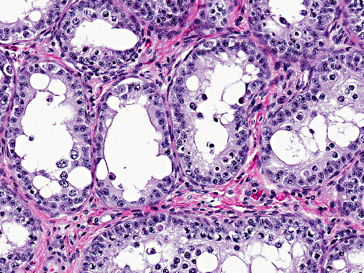
The number of seminiferous tubules is decreased (testicular hypoplasia); a variable amount of fibrous stroma separates the tubules. There are prominent fibrous septae with many plump oval fibroblast nuclei and pigmented macrophages with pale gray-blue non-polarizing granular cytoplasm (lipochrome, lipofuscin, ceroid). Interstitial (Leydig) cells are difficult to discern, and are therefore less populous than expected. Seminiferous tubules are diffusely hypocellular (hypoplasia) with diameters estimated at 120-130um. All tubules contain Sertoli cells, often with marked apical micro- and macrovacuolation (degeneration), creating a wispy cytoplasmic appearance. Approximately 95% of seminiferous tubules contain at least one spermatogonium within the basal compartment; mitotic figures are occasionally noted. Meiotic figures are rare, and only a small fraction of the expected number of spermatocytes is identified; many have swollen clear cytoplasm with vacuolated nuclei (degeneration) or are individuated with hypereosinophilic cytoplasm and round condensed or fragmented nuclei (apoptosis). Neither elongating nor mature spermatids are identified. Rare multinucleated syncytial (degenerate) spermatocytes or spermatids are noted; some are apoptotic (may not be present in all sections). Approximately 5% of the seminiferous tubules contain only Sertoli cells with frothy, vacuolated apical cytoplasm. There are multiple small to moderately sized perivascular interstitial aggregates of mature lymphocytes. Clusters of seminiferous tubules without apparent lumina are noted (cause and significance not apparent).
Morphologic Diagnosis:
Condition:
Contributor Comment:
Because the size and development of the mule testis is entirely controlled by the donkey sire (which supplies the male mules Y chromosome), it is more relevant to compare mule testes with donkey testes, but, as limited information is available for the jack, the stallion can also serve as a reference. The Y chromosome carries the Sry gene, which controls the development and differentiation of the testis, as well as the number of Sertoli cells and the length of the spermatogenic cycle.(5) The donkey Sry gene is significantly divergent from that of the stallion, and may be only partially successful in inducing and supporting testicular development in mules.(7) This is supported by the finding of a skewed sex ratio in mule foals (44 male:56 female) compared to horse foals (52.5 male:47.5 female)(reviewed in 7).Â
This testis has three features of hypoplasia: 1) it is small; 2) it has fewer seminiferous tubules than expected; and 3) the luminal diameter of the seminiferous tubules is decreased. The testis examined is about the size of that of a medium-sized dog, and is much smaller than expected for a 100kg animal. The average mule testis weighs 350g, while that of a jack weighs 750g, and that of a stallion weighs 900g.(4) Only a portion of the testis was submitted, thus the weight of this mini-mules testis was not measured. There is also a decrease in the number of tubules in this testis, possibly causing an apparent increase in the interstitial tissue. Mule testes are reported to have 73% fewer seminiferous tubules than jack testes, and the total length of mule seminiferous tubules is calculated to be only 57% of the total length of seminiferous tubules in jacks.(5) Finally, the average width of seminiferous tubules in mules is decreased; it was estimated at 120-130um in this mini-mule. Previous quantitative reports suggest that the average width of seminiferous tubules in mules is 127um and in jacks is 222um.5 It is 146um in stallions.(3)
The number of Leydig cells is decreased in this testis, as is expected with fewer differentiating germ cells failing to provide sufficient crosstalk to support an appropriate number of Leydig cells. Mules have approximately 67% of the number of Leydig cells identified in jacks, and about 40% of the number seen in stallions,(4) but mule Leydig cells are ultrastructurally normal and identical to those of jacks and stallions.(4,5,6) The smaller number of Leydig cells in mule testes is believed to reflect the failure of spermatogenesis, rather than be the cause of it. This is supported by anecdotal evidence that mules have very high libido among equids, presumably due to high levels of testosterone synthesized by the Leydig cells. Mules also typically demonstrate normal epididymal duct epithelium (a steroid-dependent phenotype).(1)
The number of Sertoli cells in this case is likely within normal limits, as is Sertoli cell function. Although the seminiferous tubular diameter is decreased compared to that seen in jacks, there is no waviness or buckling of the basement membrane, suggesting that the number of Sertoli cells is appropriate. Other references have shown that the number of Sertoli cells is equal in donkeys and mules,(5) and that they are ultrastructurally identical.(6) Additionally, lanthanum exclusion studies have shown that the blood-testis barrier is intact in mule testis.(6)
Failure of chromosomal pairing in the zygotene stage of meiotic prophase I leads to deletion of nascent germ cells by apoptosis. Apoptosis in the testis of mammals is a normal process, and is mediated by Fas on germ cells and FasL on Sertoli cells.(3) The number of germ cells undergoing apoptosis is increased after any insult, regardless of cause. It is also increased in testis with maturation arrest, as is present in mule testes.(3) Degeneration of seminiferous tubules in stallions frequently elicits a lymphocytic interstitial orchitis,(3) and that is the likely inciting cause of the inflammation seen in this case. Equine testes also commonly exhibit prominent fibrous tissue septa,(3) and the amount of interstitium in this testis may be within normal limits.
JPC Diagnosis:
1. Testis, seminiferous tubules: Hypoplasia, diffuse, severe.
2. Testis: Hemorrhage, focally extensive, severe.
Conference Comment:
References:
2. Benirschke K, Brownhill L, Beath, M: Somatic chromosomes of the horse, the donkey, and their hybrids, the mule and the hinny. J Reprod Fert 4:319-326, 1962.
3. Foster R, Ladds P: Male Genital System. In: Jubb, Kennedy, and Palmers Pathology of Domestic Animals, ed. Maxie MG, 5th ed., vol. 3, pp. 566, 572-3, 580, 585. Elsevier Limited, St. Louis, MO, 2007.
4. Hernandez-Jauregui P, Monter H: Fine structure of mule testes: light and electron microscopy study. Am J Vet Res 38:443-447, 1977.
5. Neves E, Chiarini-Garcia H, Franca L: Comparative testis morphometry and seminiferous epithelium cycle length in donkeys and mules. Biol Reprod 67:247-255, 2002.
6. Neves E, Chiarini-Garcia H, Franca L: Ultrastructural observation of the mule testis indicates normal function of somatic cells. Anim Reprod 2:263-271, 2005.
7. Short R: An introduction to mammalian interspecific hybrids. J Here'd 88:355-357, 1997.