Signalment:
10 year old neutered male domestic short hair cat (
Felis cats).The cat presented for chronic history of respiratory distress. Mass identified in trachea.
Gross Description:
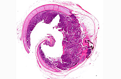
Focally extensive thickening/protrusion of the tracheal mucosa that partially occluded the lumen (1.5 cm x 1 cm x 0.25 cm).
Histopathologic Description:
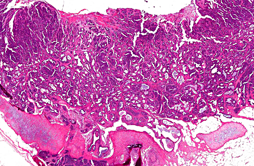
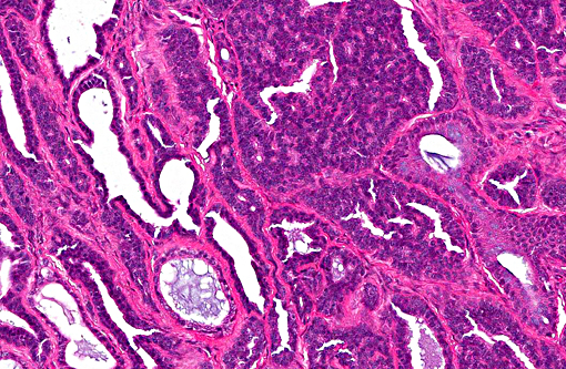
Sections of trachea in which there are marked thickening/expansion of the mucosal area by a partly delineated and nonencapsulated mass within the subepithelial/lamina propria area that contains submucosal glands. The mass is composed of cells arranged in numerous variably-sized acini and small cysts with occasional dense clusters in one area supported by a fine fibrovascular stroma. The cysts are lined by one to several layers of cells that are cuboidal with modest amounts of basophilic cytoplasm and a single oval nucleus with finely dispersed chromatin. Mitoses are not seen. Some of the acini and cysts contain small amounts of eosinophilic secretion product. Surrounding many of the cysts are minimal multifocal infiltrates of lymphocytes. A few cells of the mass extend into the subjacent tracheal cartilage in an area.
Morphologic Diagnosis:
Trachea: Mucosal adenocarcinoma
Condition:
Tracheal adenocarcinoma
Contributor Comment:
The mass appears to have arisen from glands of the tracheal mucosa and, as described, protrudes into the tracheal lumen and extends at least in one area into the subjacent tracheal cartilage. Mitotic figures are not common and there is little nuclear atypia; therefore, an adenoma is a consideration. However, the mass appears to be expansile replacing the mucosa in areas and, as indicated, extends to the tracheal cartilage. Tumors of the trachea are very rare in dogs and cats. One publication describes a tracheal tumor in a cat.(3) It is unlikely that the mass is metastatic since: a) Metastasis to the trachea is rare, and b) other neoplastic masses were not identified in the animal. Other types of neoplasms that can occur in the trachea include: Squamous cell carcinoma (especially in humans that smoke), adenoid cystic carcinoma, papilloma, chondroma, hemangioma, and carcinoid. This location of the mass has similarities to adenoid cystic carcinoma seen in humans.(5)
JPC Diagnosis:
Trachea: Mucosal adenocarcinoma.
Conference Comment:
Characteristics of this unique neoplasm which generated discussion during the conference include the focal area of infiltration into tracheal cartilage (may not be present in all slides) and the fact that it was circumferential in nature, affecting the entirety of tracheal mucosa. Participants noted frequent piling of cells and the focal invasion as two characteristics indicative of a more malignant phenotype. Most agreed the neoplasm was likely primary in the trachea due to the resemblance of the neoplasm to tracheal glands and the fact that another primary tumor was not reported in this animal.
Other primary tracheal neoplasms reported in domestic species, not listed above, include undifferentiated carcinoma, plasmacytoma, leiomyoma, fibrosarcoma, mast cell tumor, rhabdomyosarcoma, osteochondroma, osteosar-coma,(7) and primary intratracheal lymphosar-coma.(2) Epithelial tumors and osteo-chondroma are the most common tumors of the feline and canine trachea, respectively.(4) Laryngeal tumors are also considered rare, but occur more frequently than tracheal tumors. Papillomas and squamous cell carcinomas are the most common laryngeal tumors in dogs, and laryngeal lymphomas are occasionally seen in cats.(6) Neoplasms can be extraluminal or intraluminal and cause swelling in the neck and/or tracheal obstruction, respectively, depending on location; dyspnea, cough and dysphagia are common associated clinical signs. Studies have suggested an association between tobacco smoke and oral squamous cell carcinoma in cats,(1) but the precise relationship between tobacco smoke and respiratory tract tumors in cats is unclear, although it can be postulated, based on secondhand smoke being a known human carcinogen.
References:
1. Bertone ER, Snyder LA, Moore AS. Environmental and lifestyle risk factors for oral squamous cell carcinoma in domestic cats. J Vet Intern Med. 2003; 17(4):557-62.
2. Brown MR, Rogers KS, Mansell KJ, Barton C. Primary intratracheal lymphosarcoma in four cats. J Am Anim Hosp Assoc. 2003;39(5):468-472.
3. Cain GR, Manley P. Tracheal adenocarcinoma in a cat. JAVMA. 1983;182(6):614-616.
4. Carlisle CH, Biery DN, Thrall DE. Tracheal and laryngeal tumors in the dog and cat: Literature review and 13 additional patients. Vet Radiol. 1991;32:229-235.
5. Hu Z, Meng Y, Wu H, Shen J, Bi Y, Luo Y, et al. Adenoid cystic carcinoma of the tracheobronchial tree: Cliniocpathologic and immunohistochemical studies of 21 cases. Int J Clin Exp Pathol. 2014;7(100):7527-7535.
6. Lopez A. Respiratory system, mediastinum, and pleurae. In: McGavin MD, Zachary JF, eds. Pathologic Basis of Veterinary Disease. 5th ed. St. Louis, MO: Mosby Elsevier; 2012:480.
7. Ramirez GA, Altimira J, Vilafranca M. Cartilaginous tumors of the larynx and trachea in the Dog: Literature review and 10 additional cases (1995-2014). Vet Pathol. 2015; Apr 16, Epub: 1-