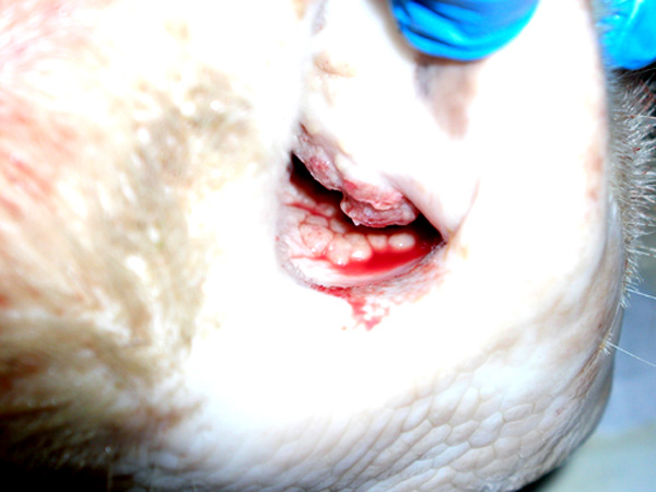
Signalment:
Gross Description:
Morphologic Diagnosis:
Lab Results:
Bacterial culture of gross lesions: Small number of contaminants (Streptococcus sp., Bacillus sp.)
BVD immunohistochemistry (ear notch): negative
Condition:
Contributor Comment:
Although uncommon, Pseudoallescheria boydii typically causes localized infections in cutaneous and subcutaneous connective tissues. Within lesions, the organism is often arranged as densely entangled hyphae (2-5_m) and swollen cells (15-25 _m) that can be grossly evident as tissue grains or granules. Within the nasal mucosa of this cow, the organisms were disseminated, and even when visualized with silver stains, did not form entangled hyphae. In fact, hyphae were inconspicuous compared to the variably-sized spherical swollen cells.Â
Other than the cutaneous and subcutaneous mycetomas, Pseudoallescheria boydii has also been implicated in bovine abortions.
JPC Diagnosis:
Conference Comment:
P. boydii are ubiquitous within the environment. However, infections by this fungus are extremely rare and primarily reported in immunocompromised patients. In this case, there was no evidence the cow was immunocompromised. In animals, P. boydii primarily causes trauma-induced eumycotic mycetomas. It has rarely been associated with equine and bovine abortions, pneumonia in a calf, granulomatous rhinitis and onychomycosis in the horse, and eumycotic mycetoma and keratomycosis in the dog and horse. Unlike in dogs, nasal infections of cattle with P. boydii do not typically invade the underlying bone.Â
Gross differentials for rhinitis in cattle include atopic rhinitis, neoplasia (e.g. lymphoma, squamous cell carcinoma), foreign body, actinobacilloisis, actinomycosis, and other fungal diseases (e.g. rhinosporidiosis, aspergillosis and phycomycosis). P. boydii differs from Aspergillus and Fusarium sp. by an absence of both angioinvasion and dichotomous branching.
Treatment of P. boydii is difficult and requires antifungal-susceptibility testing since the organism exhibits some level of inherent resistance to most antifungal agents.
References:
2. Hargis AM, Ginn PE: The integument. In: Pathologic Basis of Veterinary Disease, eds. McGavin MD, Zachary JF, 4th ed., pp. 1194-1195. Mosby Elsevier, St. Louis, Missouri, 2007
3. Knudtson WU, Kirkbride CA: Fungi associated with bovine abortion in the northern plains states (USA). J Vet Diagn Invest 4:181-185, 1992
4. Kwon-Chung KJ, Bennett JE: Pseudallescheriasis and Scedosporium infection. In: Medical Mycology, pp. 678-694. Lea & Febiger, Malvern, PA, 1992
5. Larone DH: Medically Important Fungi: A Guide to Identification, 4th ed., pp. 196-197. ASM Press, Washington, DC, 2002
6. Rippon JW: Medical Mycology: The Pathogenic Fungi and the Pathogenic Actinomycetes, 3rd ed., pp. 651-677. W. B. Saunders Company, Philadelphia, PA, 1988
7. Schlafer DH, Miller RB: Female genital system. In: Jubb, Kennedy, and Palmers Pathology of Domestic Animals, ed. Maxie MG, 5th ed., vol. 3, pp. 508-509. Elsevier Limited, St. Louis, MO, 2007
8. Schwartz DA: Pseudallescheriasis and Scedosporiasis. In: Pathology of Infectious Diseases, eds. Connor DH, Chandler FW, Schwartz DA, Manz HJ, Lack EE, vol. 2, pp. 1073-1079. Appleton & Lange, Stamford, CT, 1997
9. Singh K, Boileau MJ, Streeter RN, Welsh RD, Meier WA, Ritchey JW: Granulomatous and eosinophilic rhinitis in a cow caused by Pseudallescheria boydii species complex (anamorph Scedosporium apisospermum). Vet Path 44:917-920, 2007
