Conference 23
CASE III:
Signalment:
1-year-old, intact male (stud colt), American Quarter Horse, Equus caballus, horse.
History:
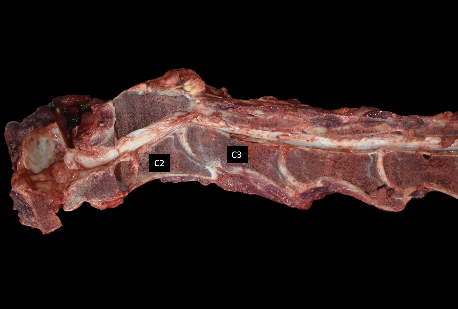
The animal was presented to the large animal teaching hospital for progressive lameness of the hind limbs and a possible pelvic injury after a collision with another horse in pasture six months prior resulted in a fall. Lameness examination revealed a marked grade three lameness of the left hind limb and limited flexion of the stifles with both hind toes worn down from dragging. On neurologic examination, he circumducted his outside hind limb when turning in tight circles. When his head was lifted and he was walked forward, his forelimbs hovered for a prolonged period before he placed them back on the ground. He had mild difficulty backing up and stumbled when walking down the grass hill. Radiographs of the cervical spinal cord revealed severe dorsal deviation of C3 relative to the caudal endplate of C2 and severe stenos is of the spinal canal at C2-C3. The owners elected for euthanasia and an autopsy was performed the same day.
Gross Pathology:
The 2nd and 3rd cervical vertebral joints were stiff and fixed in a flexed position with minimal manual flexion available. The flexed position resulted in a narrow cervical spinal canal at the level of the 2nd and 3rd cervical vertebrae with dorsal compression and flattening of the underlying spinal cord.
Laboratory Results: N/A
Microscopic Description:
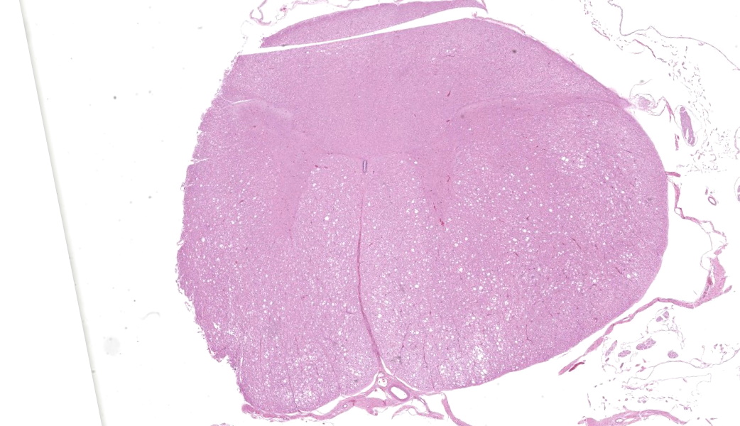
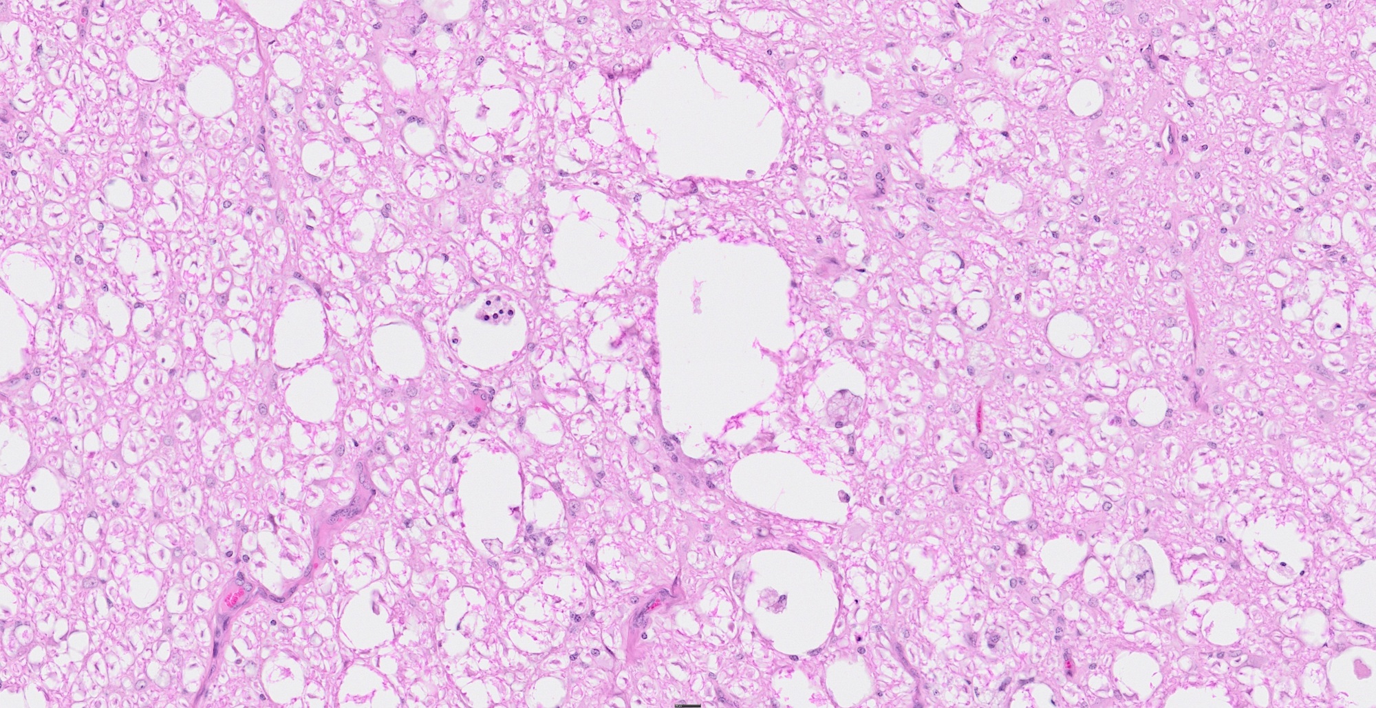
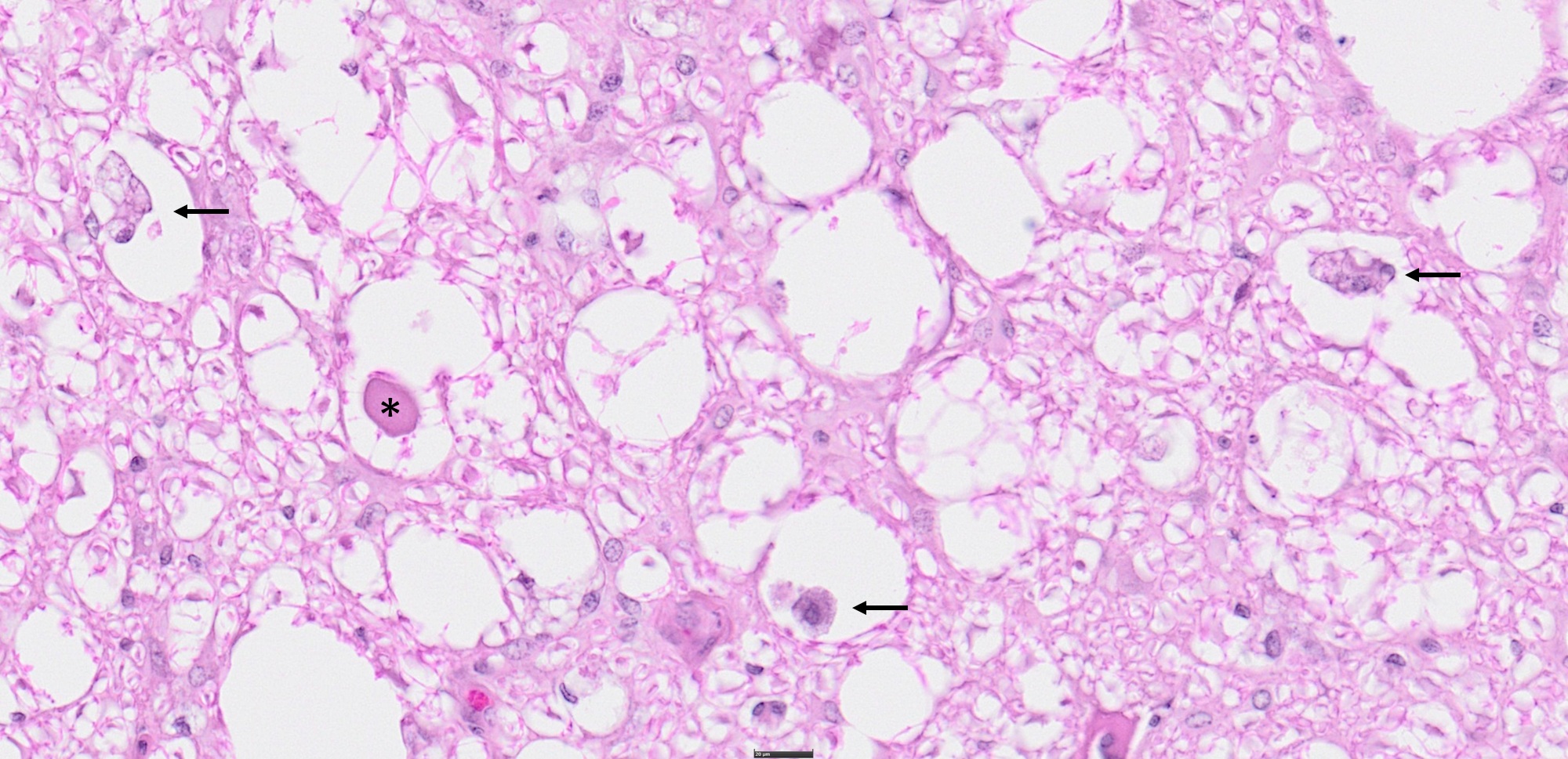
Cervical spinal cord: Most severely affecting the white matter of the ventral and lateral funiculi, and to a lesser extent the dorsal funiculus, is extensive axonal degeneration in which 60% of myelin sheaths are markedly dilated and occasionally coalesce to form up to 80-um diameter clear spaces. The dilated myelin sheaths frequently contain swollen, eosinophilic axons (spheroids) or fragments of cellular debris with occasional axonophages (digestion chambers).
Contributor's Morphologic Diagnosis:
Cervical spinal cord: Axonal degeneration, locally extensive, marked, chronic withspheroids and digestion chambers (Wallerian degeneration).
Contributor's Comment:
The clinical course of the disease paired with the gross and histopathologic findings are consistent with cervical vertebral stenotic myelopathy (CVSM) with static stenosis. CVSM (also called cervical vertebral malformation-malarticulation or colloquially referred to and cited in older text as wobbler syndrome) is a common cause of spinal cord compression in horses and dogs.1,4,5 The condition is characterized by a stenotic cervical vertebral canal and secondary extramedullary compression of the cervical spinal cord.1,2,4 Microscopically, lesions within the affected spinal cord are typical of compressive injury to the spinal cord with extensive axonal degeneration leading to demyelination at the compression site.1,4 White matter degeneration rostral to the compression is typically limited to the ascending tracts of the dorsal funiculi, while caudal is limited to the descending tracts of the ventral funiculi.1
In horses, two manifestations of the condition include cervical vertebral instability (dynamic stenosis) and cervical vertebral malformation (static stenosis). Dynamic compression occurs intermittently when the neck is flexed and typically occurs at C3-C5 vertebrae in horses 8 to 18 months of age.1,4,5 In contrast, static stenosis is a constant compression regardless of neck position and typically occurs in horses 1-4 years of age and older, with dorsal or dorsolateral narrowing of the canal most commonly at C5-C7 vertebrae.1,4 Lesions thought to contribute to static stenosis include thickening of the ligamentum flavum, thickening of the dorsal lamina, or osteophytosis or osteochondrosis of the articular processes.4,5 Bone cysts of
the articular processes have also been reported, but further investigation is needed to determine their significance and correlation/causation.2 The time required for development of lesions may account for the older age of onset seen with static stenosis. For this case, changes to the articular facets were not appreciated and a section of the ligamentum flavum was histologically unremarkable.
The pathogenesis of CVSM remains incompletely understood and is thought to be multifactorial with proposed pathways including disordered bone and cartilage maturation or abnormal biomechanical forces and altered vertebral structure.2,5 Predisposing factors are thought to include rapid growth rates, nutritional over supplementation, and genetic predisposition.1,2,4 Males and large breeds (thoroughbreds, Tennessee Walking Horses, and Warmbloods) have been shown to be at greatest risk for development.1,3,4
An interesting component of this case history is the worsening of lameness immediately following a traumatic incident (collision with another horse and fall). As the severity of CVSM is considered dependent on the speed and extent of spinal cord compression, one can speculate that the incident likely induced rapid compression of the damaged axons and secondary damage that the tissue was unable to accommodate for, in contrast to the slowly progressive compression induced by the static stenosis alone.1
Contributing Institution:
166 Greene Hall College of Veterinary Medicine Auburn University, AL 36849 https://www.vetmed.auburn.edu/academic-departments/deptof-pathobiology/
JPC Morphologic Diagnosis:
Cervical spinal cord, ventral and ventrolateral funiculi: Axonal degeneration and loss, subacute, regionally extensive, severe.
JPC Comment:
This case provided an excellent opportunity to revisit the neuroanatomic significance behind cervical vertebral stenotic myelopathy (CVSM) and the characteristic ventral white matter degeneration that results from chronic spinal cord compression. This case was classic for this disease in almost all aspects. The clinical presentation and provided imaging
correlated beautifully with the histologic pattern of ventral funiculus-predominant Wallerian-like degeneration, with spheroids, digestion chambers, and myelin loss. Many thanks to our contributor for providing fantastic supplemental images!
Participants reviewed the organization of spinal cord tracts, noting that descending motor pathways-including the lateral and ventral corticospinal tracts, reticulospinal tracts, and vestibulospinal tracts-are concentrated in the ventral and ventrolateral funiculi. In contrast, the dorsal and dorsolateral funiculi are dominated by ascending sensory tracts. This distinction is crucial because a lesion that selectively affects ventral white matter is almost always a descending motor tract problem. In horses, the spinal cord compression that occurs in this condition results in the proprioceptive ataxia that earns the condition the colloquial name of "Wobbler's disease."6 The contributor provides a well-written overview of the two manifestations of this syndrome in horses.1,4,6
The age and breed in this case were somewhat atypical, prompting discussion about how trauma, growth rate, genetics, and vertebral conformation can result in differences in the expected presentation.2 Regardless of the initiating factors, the histologic result is the same: chronic compression leading to Wallerian-like axonal degeneration.
Dr. Koehler highlighted the distinction between Wallerian degeneration and Wallerian-like degeneration, a nuance that often confuses trainees. True Wallerian degeneration was originally defined in the context of acute, severe, transecting injury to the spinal cord, where axons undergo rapid fragmentation distal to the site of injury.2 In contrast, Wallerian-like degeneration describes the same morphologic process, but arising from chronic, progressive insults such as compression, ischemia, or metabolic injury.2 Histologically, the two are nearly indistinguishable; the difference lies in the tempo and mechanism of axonal injury. In this case, the chronicity of the lesion supports the use of the term "Wallerian-like". Similar to case 1, the JPC's morph reflects the participants' view that axonal degeneration and loss are the primary lesions in this case and encompass many of the other findings, including spheroids, digestion chambers, and demyelination since these are all considered manifestations of or sequalae to axonal degeneration/loss.
References:
- Craig LE, Dittmer KE, Thompson KG. Nervous System. In: Zachary JF. Pathologic Basis of Veterinary Disease. St. Louis, MO: Elsevier; 2021: 952-954
- Janes JG, et al. Cervical vertebral lesions in equine stenotic myelopathy. Veterinary Pathol. 2015; 52:919-27.
- Levine JM, Ngheim PP, Levine GJ, Cohen, ND. Associations of sex, breed, and age with cervical vertebral compressive myelopathy in horses: 811 cases (1974-2007). Journal of the American Veterinary Medical Association. 2018; 233: 1453-1458.
- Miller AD, Porter BF. Developmental Diseases of Joints. In: Maxie, MG. Jubb, Kennedy, and Palmer's Pathology of Domestic Animals. 6th ed, vol. 1. St. Louis, MO: Elsevier; 2016: 136.
- Powers BE, Stashak TS, Nixon AJ, Yovich JV, Norrdin RW. Pathology of the Vertebral Column of Horses
- Woodie B, Johnson AL, Grant B. Cervical Vertebral Stenotic Myelopathy. Vet Clin North Am Equine Pract. 2022;38(2):225-248.