Signalment:
Gross Description:
Histopathologic Description:
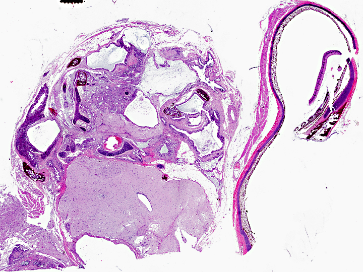
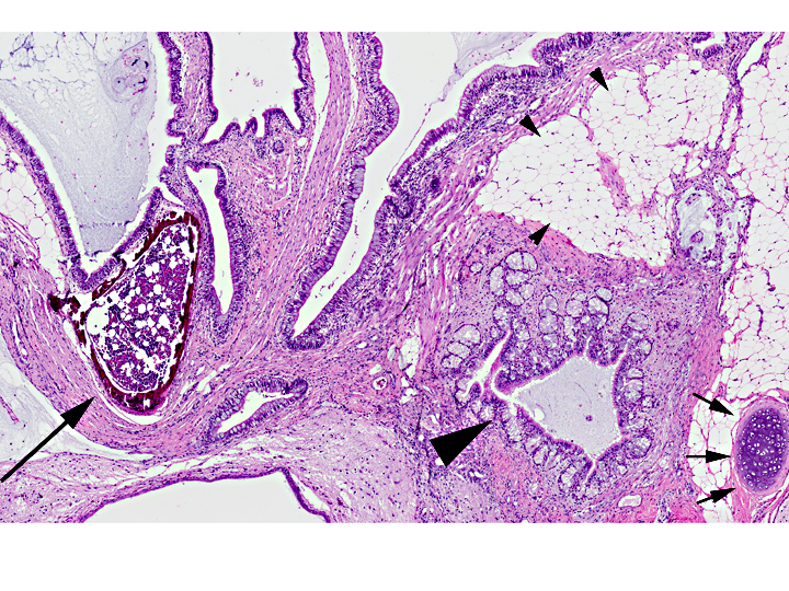
Multifocally, solid areas are composed of moderately cellular neoplastic neural tissue with neurons and glial cells embedded in neuropil (ectoderm). There are multifocal areas of formation of bone with marrow and cartilage (mesoderm). In a few of the sections, there are small lobules of zymogen-filled acini, resembling pancreas (endoderm). Mitoses in all cell populations are not prominent (fewer than 1 per HPF). In the small area of eye present, there is evidence of retinal detachment, hypertrophy of the retinal pigment epithelium, and retinal degeneration.
Morphologic Diagnosis:
Condition:
Contributor Comment:
They are uncommon in domestic animals, but have been reported mostly in the bitch, sow, mare and cow; their most common localization is the gonads.(7,11) In avian species, involvement of the testes appears to be more common than the ovary9 and they are most commonly observed in chickens.(6,10)
There are two reports of retrobulbar teratomas in wild birds.(6,10) One of the tumors was poorly differentiated and interpreted as malignant.(6) Incidentally, old literature reports describe experimental induction of teratomas in testicles of fowl by local injection of metal ions (zinc) in young animals.(2,3,9), which was initially intended as a method of chemical castration.(2,3)
Teratomas in animals, in contrast to those in humans, are almost always benign.(7) In humans, classification of a teratoma as benign or malignant is based largely on the identification of primitive, undifferentiated cells and tissues within the tumor.(1) In general, the presence of a germ cell component worsens the prognosis, and teratomas with incompletely differentiated tissues should be considered potentially malignant.(1,5)
In this particular case, the eye was submitted as an enucleation and the kestrel was presumed alive at the time of biopsy. Unfortunately, follow-up evaluation was not available.
JPC Diagnosis:
Conference Comment:
Several conference participants were unable to identify ectodermal differentiation to haired skin due to slide variability; however, this does not affect the diagnosis of teratoma, since only 2 germ cell lines are required to make the diagnosis. Although most reports define teratomas as having at least 2 of the 3 germ cell lines, rare variants (in humans) have only 1 line (monodermal variant).
References:
2. Guthrie J. Specificity of the metallic ion in the experimental induction of teratomas in fowl. Br J Cancer. 1967;21:619-22.
3. Guthrie J. Zinc induction of testicular teratomas in Japanese quail (Coturnix coturnix Japonica) after photoperiodic stimulation of testis. Br J Cancer. 1971;25:311-4.
4. Hooper CC. Teratoma in the cerebrum of a fantail pigeon. Avian Pathol. 2008;37:141-3.
5. Klein MK. Tumors of the female reproductive system. Germ cell tumors. In: Withrow SJ, MacEwen EG, eds. Small Animal Clinical Oncology. 3rd edition. St. Louis, MO: Saunders; 2001:446.
6. L³pez RM, M+â°rcia DB. First description of malignant retrobulbar and intracranial teratoma in a lesser kestrel (Falco naumanni). Avian Pathol. 2008;37:413-4.
7. MacLachlan NJ, Kennedy PC. Tumors of the genital systems, Teratoma. In: Meuten DJ ed. Tumors in Domestic Animals. 4th edition. Ames, Iowa: Iowa State University Press, 2002:554.
8. Petrak ML, Gilmore CE. Neoplasms. In: Petrak ML, ed. Diseases of Caged and Aviary Birds. 2nd ed. Philadelphia, PA: Lea and Febiger; 1982:606-637.
9. Reece RL. Tumors of unknown etiology. In: Calnek BW, ed. Disease of Poultry. 10th ed. Ames, Iowa: Iowa State University Press, 1997:496-497.
10. Schelling SH. Retrobulbar teratoma in a great blue heron (Ardea herodias). J Vet Diagn Invest. 1994;6:514-6.
11. Shlafer DH, Miller RB. Female genital system. In: Maxie MG, ed. Jubb, Kennedy, and Palmers Pathology of Domestic Animals. 5th edition. Vol 3. St. Louis, MO; Saunders Elsevier; 2007:453-454.
12. Stricker TP, Kumar V. Neoplasia. In: Kumar V, Abbas AK, Fausto N, Aster JC, eds. Robbins and Cotran Pathologic Basis of Disease. 8th ed. Philadelphia, PA: Saunders Elsevier; 2010:261-2.