Signalment:
membranes. All lymph nodes were slightly prominent. Radiograph showed an enlarged, pendulous abdomen with hepatomegaly and severe splenomegaly. The ultrasound examination revealed that the liver is covered with hypo echoic, nodule-like structures. The visible mesenteric lymph nodes were enlarged. Laboratory tests: Coagulation time was significantly prolonged. Liver-associated values were as well significantly altered. Because of poor prognosis the animal was euthanized.
Gross Description:
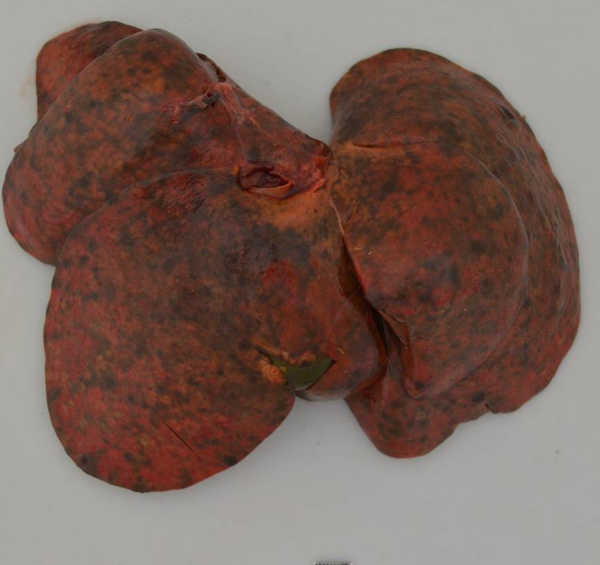
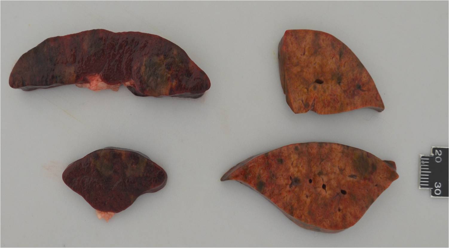
Liver: Diffusely severely enlarged, with disseminated white-green and black, up to 5mm nodules which are multifocally slightly raised and show central necrosis.
Spleen: Diffusely severely enlarged. Multifocally are up to 4 cm nodules present that are dark red and slightly firm.
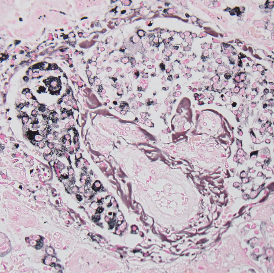
Histopathologic Description:
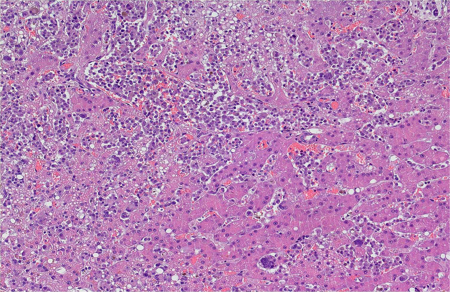
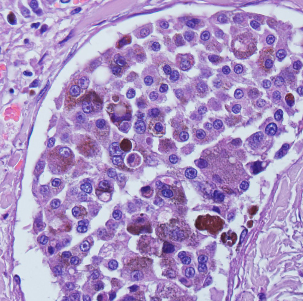
Masson-Fontana: Many of the neoplastic cells contain variable amounts of intra-cytoplasmatic, agyrophilic, granular material (melanin).
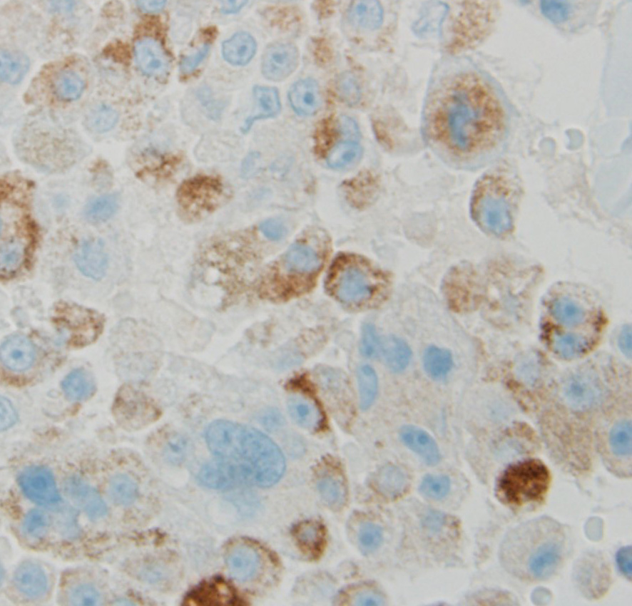
Immunohistochemistry: The neoplastic cells stain positive with -Ç-ÿMelan A and -Ç-ÿPNL-2 (Melanoma).
Morphologic Diagnosis:
Lab Results:
Condition:
Contributor Comment:
Malignant melanomas are generally tumors of older animals; however, they have been reported in juvenile animals of many species.2 They are common in the dog and uncommon in other domestic species. In cats, they are uncommon and occur mainly in older cats showing no sex predisposition. Malignant melanomas can be composed of a variety of cell morphologies including spindle cells, epithelioid cells, a mixture of spindle cells and epithelioid cells, singet-ring cells or balloon cells. In addition they can be heavily pigmented or amelanotic.3 The present metastatic mass in liver and spleen is considered as an epithelioid type and is pigmented. Metastasis occurs commonly with spread via lymphatics to regional lymph nodes and lungs. It is not uncommon for malignant melanomas to spread to other body sites, including unusual locations such as the brain, heart and spleen.3
Ocular melanoma in cats occurs in a variety of sites. The vast majority of feline ocular melanomas arise diffusely from the anterior uvea (iris, ciliary body).5 Conjunctival melanomas in dogs and cats are in general rare.3 One study reported in a comparison study of 19 feline ocular melanomas that 16 were intraocular, while only three were conjunctival.4 In another study by Schobert et al. (2010), in 13 cases of conjunctival melanoma in cats with adequate follow-up information, only three reported metastasis (14%). Of those three cases, two had metastasized to the submandibular lymph nodes, while in the third case, an abdominal mass was detected.5 Interesting about our case is that two abdominal organs (liver, spleen) were severely infiltrated and that the neoplastic cells were present in the lymphatics and blood vessels diffusely within the spleen and liver.
JPC Diagnosis:
Conference Comment:
This was a challenging case
for conference participants with the majority diagnosing this lesion as histiocytic
sarcoma due in large part to the presence of multinucleated cells.
Cholestasis, as seen in this case, is also a common feature of hepatic
histiocytic sarcoma. The moderator commented this is an unusual case and he had
not previously seen a case like this one.
Additional features described and discussed included vascular thrombosis
and, interestingly, rare ![]() mitoses were noted in hepatocytes in the absence of regenerative
nodules. The moderator pointed out the collar of connective tissue surrounding
the central vein, which is a normal feature in the feline liver that
distinguishes it from other species. The areas of hepatocyte necrosis and loss
surrounding the central vein are presumed secondary to hypoxia from obstruction
of sinusoids by tumor emboli. The excellent gross image and the laboratory
data provided by the contributor greatly enhanced the learning / teaching value
of this case.
mitoses were noted in hepatocytes in the absence of regenerative
nodules. The moderator pointed out the collar of connective tissue surrounding
the central vein, which is a normal feature in the feline liver that
distinguishes it from other species. The areas of hepatocyte necrosis and loss
surrounding the central vein are presumed secondary to hypoxia from obstruction
of sinusoids by tumor emboli. The excellent gross image and the laboratory
data provided by the contributor greatly enhanced the learning / teaching value
of this case.
References:
1. Chamel G, Abadie J, Albaric O, Labrut S. Non-ocular melanomas in cats: a retrospective study of 30 cases. J Feline Med Surg. 2016; Jan 14(E pub):1-7.
2. Ginn PE, Mansell JEKL, Rakich PM. Skin and appendages. In: Maxie MG, ed. Jubb, Kennedy, and Palmers Pathology of Domestic Animals. 5th ed. Vol 1. Philadelphia, PA: Elsevier Saunders; 2007:760.
3. Meuten DJ. Tumors in Domestic Animals. 4th ed. Iowa State Press; 2002.
4. Patnaik AK, Mooney S. Feline melanoma: a comparative study of ocular, oral, and dermal neoplasms. Vet Pathol. 1988; 25: 105-112.
5. Schobert CS, Labelle P, Dubielzig RR. Feline conjunctival melanoma: histo-pathological characteristics and clinical outcomes. Veterinary Ophthalmology. 2010;