Signalment:
Gross Description:
Histopathologic Description:
Morphologic Diagnosis:
Lab Results:
-�-� Mycoplasma hyopneumoniae PCR: NEGATIVE
-�-� Mycoplasma hyopneumoniae IHC: NEGATIVE
Condition:
Contributor Comment:
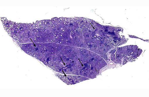
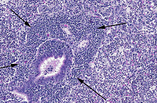
Gross lesions associated with mycoplasmal pneumonia are discolored, collapsed, firm lungs affecting the cranioventral lung lobes.(1) Acute histologic lesions are characterized by alveoli containing macrophages and neutrophils along with edema.(1) Chronic infections in swine are similar to other species (e.g., rats, mice) where peribronchial/peribronchiolar (BALT), and perivascular lymphoid hyperplasia is a dominant histologic feature.
Although the exact pathogenesis is not completely understood, Mycoplasma hyopneumoniae firmly adheres to the cilia of the respiratory tree resulting in ciliostasis. Attachment to the respiratory epithelium invokes the following:
(1) Influx of neutrophils into the tracheobronchial mucosa
(2) Extensive loss of cilia (deciliation)
(3) Broncho-alveolar lymphoid tissue (BALT) hyperplasia
(4) Influx of mononuclear cells into the peribronchiolar, bronchiolar, and alveolar interstitium(3)
Diagnosis of Mycoplasma hyopneumoniae infection in the porcine lung is based on results of three methods: isolation of the organism by culture, immunofluorescence (IF) testing, or immunohistochemistry using polyclonal antibodies.(3)
Despite our inability to definitively diagnose Mycoplasma hyopneumoniae as a causative agent for the lung lesions in this case, the histologic lesions are highly suggestive of this entity.
JPC Diagnosis:
Conference Comment:
The contributor provides an excellent summary of M. hyopneumoniae. See WSC 2013-2014, conference 7, case 1 for further discussion of Mycoplasma spp., and table 1 for an abbreviated list of Mycoplasma species important in veterinary medicine.
Table 1. Select Mycoplasma species of veterinary importance.4
| Mycoplasma species | Hosts | Disease |
| M. mycoides subsp. mycoides (small colony type) | Bovine | Contagious bovine pleuropneumonia |
| M. bovis | Bovine | Mastitis, pneumonia, arthritis, otitis |
| M. agalactiae | Ovine, Caprine | Contagious agalactia (mastitis) |
| M. capricolum subsp. capripneumoniae | Caprine | Contagious caprine pleuropneumonia |
| M. capricolum subsp. capricolum | Ovine, Caprine | Septicemia, mastitis, polyarthritis, pneumonia |
| M. mycoides subsp. capri (includes strains previously classified as M. mycoides mycoides large colony type) | Ovine, Caprine | Septicemia, pleuropneumonia, mastitis, arthritis |
| M. ovipneumoniae | Ovine, Caprine | Pneumonia |
| M. pulmonis | Rodents--rat and mouse | Colonize nasopharynx and middle ear; affect respiratory and reproductive tracts and joints |
| M. hyopneumoniae | Swine | Enzootic pneumonia |
| M. hyosynoviae | Swine (10-30 weeks of age) | Polyarthritis |
| M. hyorhinis | Swine (3-10 weeks of age) | Polyserositis |
| M. suis | Swine | Mild anemia, poor growth rates |
| M. ovipneumoniae | mild pneumonia | |
| M. haemofelis | Feline | Feline infectious anemia |
| M. cynos | Canine | Implicated in kennel cough complex |
| M. haemocanis | Canine | Mild or subclinical anemia; more severe signs in splenectomized animals |
| M. gallisepticum | Turkeys and Chickens | Chronic respiratory disease; infectious sinusitis |
| M. synoviae | Turkeys and Chickens | Infectious synovitis |
| M. meleagridis | Feline, Equine | Conjunctivitis in cats, pleuritis in horses |
| M. equigenitalium | Equine | Abortion |
References:
1. Caswell JL, Williams KJ. Respiratory system. In: Maxie MG, ed. Jubb, Kennedy, and Palmers Pathology of Domestic Animals. Vol 2. 5th ed. Vol. 2. Philadelphia, PA: Elsevier Saunders; 2007;591-592.
2. Kwon D, Chae C. Detection and localization of Mycoplasma hyopneumoniae DNA in lungs from naturally infected pigs by in situ hybridization using a digoxigenin-labeled probe. Vet Pathol. 1999;36:308-313.
3. Lopez A. Respiratory system, mediastinum, and pleurae. In: Zachary JF, McGavin MD, eds. Pathologic Basis of Veterinary Disease. 5th ed. St. Louis, MO: Elsevier Mosby; 2012:494-504, 520-521.
4. Quinn PJ, Markey BK, Leonard FC, FitzPatrick ES, Fanning S, Hartigan PJ. Mycoplasmas. In: Veterinary Microbiology and Microbial Disease. 2nd ed. Ames, Iowa: Wiley-Blackwell; 2011; Kindle edition.