Signalment:
Gross Description:
Histopathologic Description:
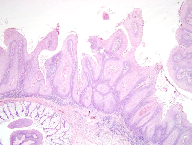
Cloaca: The normal epithelium is replaced by abundant papillary structures. The papillary structures are supported by a fibrovascular stroma and lined by markedly hyperplastic stratified squamous epithelium (Fig. 4-1). There is marked parakeratotic hyperkeratosis. There are an increased number of mitotitic figures among the basal epithelial cells (36 mitotic figures/10hpf). Between the folds of the papillary structures and extending along the surface is necrotic cellular debris, and aggregates of cocci bacteria. Within the fibrous stroma there is a mild diffuse mixed inflammatory infiltrate consisting of macrophages, plasma cells, heterophils and lymphocytes.
The second section of cloaca: There is mild erosion of the overlying mucosal epithelium.
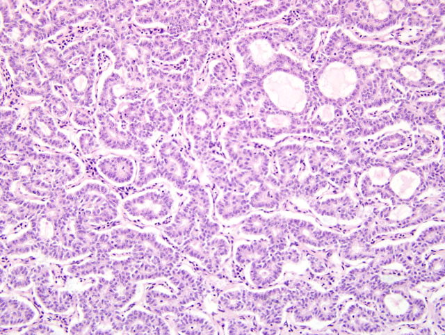
Liver: There is a focally extensive mass with an invasive pattern of growth at the margins replacing the normal hepatic architecture. The mass consists of pleomorphic cells forming tubular structures supported by a fine fibrovascular stroma (Fig. 4-2). The cells vary from columnar to cuboidal, have a moderate to scant amount of eosinophilic cytoplasm, oval to round nucleus, reticular chromatin and prominent nucleolus. There is moderate anisokaryosis and increased nuclear to cytoplasmic ratio. Mitotic figures are rare. There are multiple small aggregates of lymphocytes within the mass.Â
Morphologic Diagnosis:
Liver: Bile duct carcinoma
Lab Results:
Condition:
Contributor Comment:
Recent studies have demonstrated an alphaherpesvirus, within cloacal, oral and cutaneous papillomatous lesions and normal cloacal tissue.4,6 An alphaherpesvirus was identified by PCR analysis of cloacal tissue in the scarlet macaw presented in this case. Styles D and co-workers demonstrated the virus isolated from cutaneous & cloacal papillomas and the normal cloacal mucosa of African grey parrots (Psittacus erithacus erithacus) was most closely related, phylogenetically, to the psittacid herpesvirus (which causes Pachecos disease, psittacid herpesvirus 1), but demonstrated sufficient nucleotide and amino acid diversity to be considered a new alphaherpesvirus, psittacid herpesvirus 2.6
Pachecos disease is a devastating disease with acute onset. Histopathological findings include marked hepatic necrosis, with intranuclear inclusion bodies. Splenic necrosis, enteritis, pancreatitis, tracheitis and air sacculitis are other lesions which are seen variably.1
JPC Diagnosis:
2. Cloaca: Papilloma.Â
Conference Comment:
Psittacid herpesvirus-2 DNA sequence differs from psittacid herpesvirus-1, the cause of Pacheco's disease, by more than 20%.8 All four psittacid herpesvirus-1 geneotypes have been shown to cause Pacheco's disease in Amazon parrots, but only genotypes 2, 3 and 4 result in disease in African grey parrots.8
Other causes of cloacal papillomas or papilloma-like lesions include: papillomavirus; chronic irritation with cell hypertrophy or hyperplasia; and malnutrition with vitamin A deficiency.2 Papillomavirus infections in birds have been demonstrated in an African grey parrot, finch, and Cuban Amazon parrot.3,6
References:
2. Gerlach H: Viruses. In: Avian Medicine: Principle and Applications, eds. Ritchie BW, Harrison GJ, Harrison LR, pp. 886-888. Wingers Publishing Inc., Lake Worth, FL, 1994
3. Gibbons PM, Busch MD, Tell LA, Graham JE, Lowenstine LJ: Internal papillomatosis with intrahepatic cholangiocarcinoma and gastrointestinal adenocarcinoma in a peach-fronted conure (Aratinga aurea). Avian Dis 46:1062-1069, 2002
4. Johne R, Konrath A, Krautwald-Junghanns ME, Kaleta ER, Gerlach H, Muller H: Herpesviral, but no papovaviral sequences, are detected in cloacal papillomas of parrots. Arch Virol 147:1869-1880, 2002
5. Latimer KS: Oncology. In: Avian Medicine: Principle and Applications, eds. Ritchie BW, Harrison GJ, Harrison LR, pp. 657-658. Wingers Publishing Inc., Lake Worth, FL, 1994
6. Styles DK, Tomaszewski EK, Phalen DN: A novel psittacid herpesvirus found in African grey parrots (Psittacus erithacus erithacus) Avian Pathol 34:150-154, 2005
7. Tomaszewski EK, Kaleta EF, Phalen DN: Molecular phylogeny of the psittacid herpesviruses causing Pacheco's disease: Correlation of genotype with phenotypic expression. J Virol 77:11260-11267, 2003
8. Tomaszewski EK, Wigle W, Phalen DN: Tissue distribution of psittacid herpesviruses in latently infected parrots, repeated sampling of latently infected parrots and prevalence of latency in parrots submitted for necropsy. J Vet Diagn Invest 18:536-544, 2006