CASE II:
Signalment:
12 year-old, female, Bouvier des Flandres dog (Canis lupis familiaris)
History:
Straining to defecate for several days. Rectal palpation revealed a 1.5 cm nodule just inside the anus. The mass was surgically excised and submitted for histopathologic examination.
Gross Pathology:
Received a 1.8 x 1.5 x 0.8 cm brown tissue. The surgical border could not be identified at the time of sectioning.
Laboratory Results:
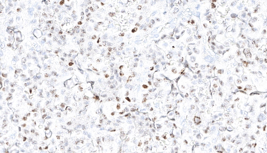
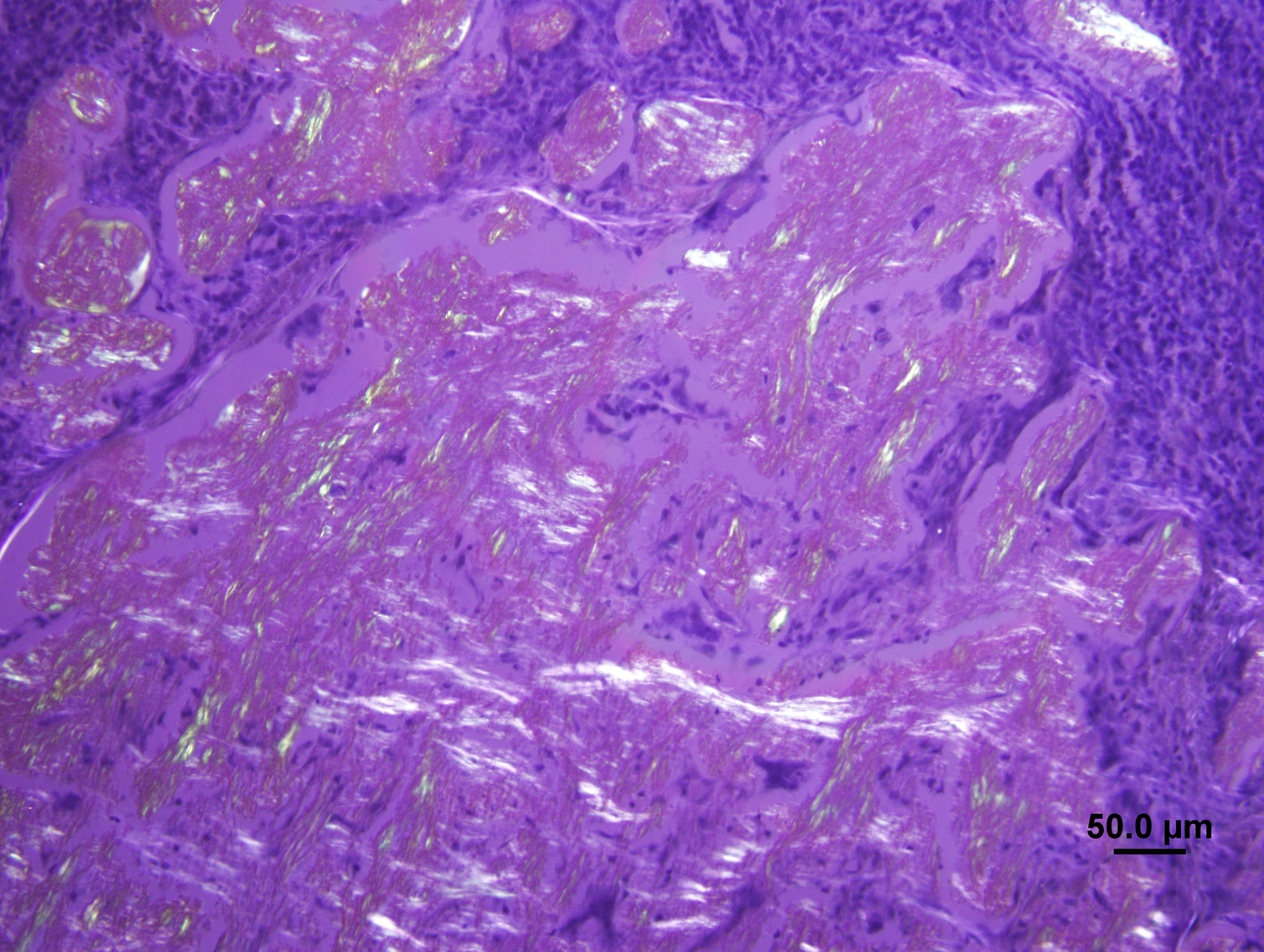
In formalin-fixed, paraffin- embedded sections of the rectal mass the tumor cells exhibited positive pyronophilia with methyl green pyronin. The deposits of amyloid stained with Congo red and exhibited some apple-green birefringence under polarized light. The birefringence was resistant to pretreatment with potassium permanganate supporting immunoglobulin-derived amyloid (primary) composed of lambda light chains. The tumor cells were immunohistochemically positive for lambda light chains.
Microscopic Description:
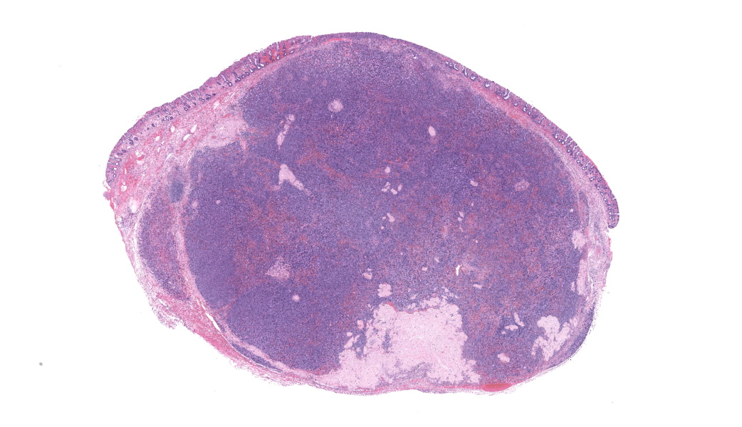
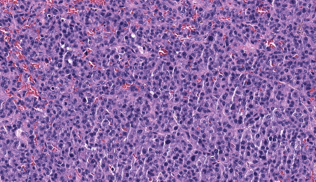
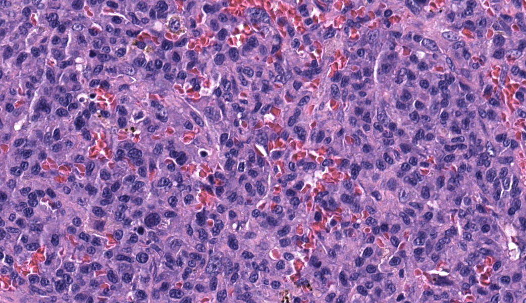
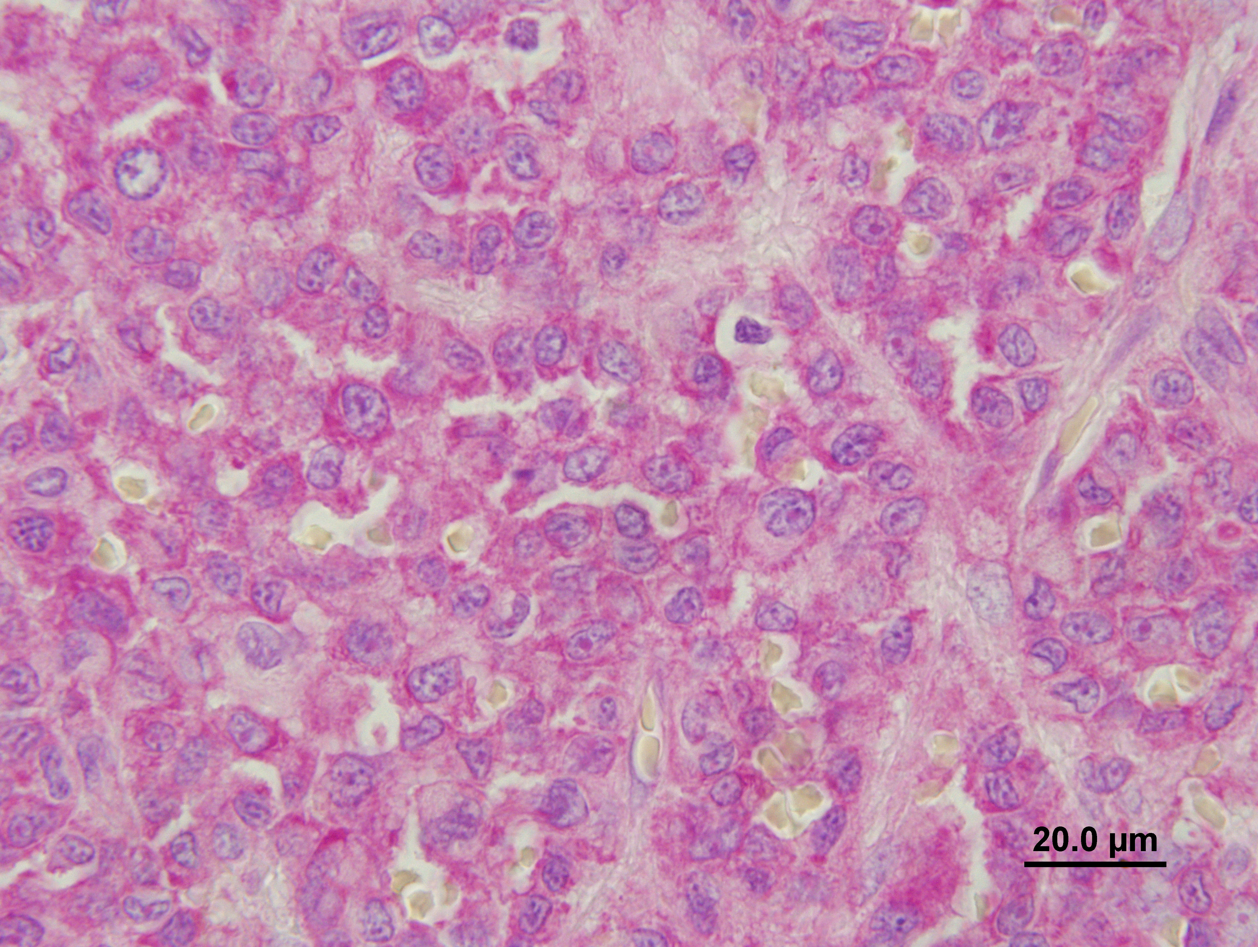
In the submucosa there is a poorly demarcated, compressive, unencapsulated mass that comprises packets, supported by thin strands of fibrovascular connective tissue, and sheets of round cells with distinct cell borders and a moderate to high nuclear to cytoplasmic ratio. Tumor cells are close to or at all of the circumferential surgical borders. The nucleus is eccentric, round to oval to indented to bizarre (lobulated forms seen) with a finely to coarsely stippled chromatin pattern. Occasional cells possess one to two, large, round nucleoli. The cytoplasm is scant to moderate and blue. Many cells have a paranuclear clear zone and some cells possess a few large, pink globules. Some cells appear to contain a coarsely granular, golden brown pigment (hemosiderin?). Anisokaryosis is moderate. Multinucleate forms (up to 3 nuclei seen) are rare. Karyomegaly is frequent. Mitoses are seen (0-2 per 40X objective fields). There are several foci of hemorrhage accompanied by a few hemosiderophages in the mass.There are extracellular collections of a pale pink, homogeneous material that is interpreted to be amyloid. The mucosa is multifocally ulcerated and there is a moderate infiltrate of neutrophils, small lymphocytes and plasma cells in the lamina propria. Hemorrhage is present in the lamina propria and submucosa.
Contributor's Morphologic Diagnosis:
Rectal mass: Colonic extramedullary plasmacytoma (EMP) with intralesional primary amyloid deposition
Contributor's Comment:
The incidence of rectal and colonic EMPs has been reported to be 4.0% of all canine EMPs2. Other reported locations include cutaneous sites (86%; mostly on the skin of the limbs, and head); the mucus membranes of the oral cavity and lips (9%) and other sites (1%; stomach, small intestine, genital system and spleen)2. Tumors occur frequently in, at or near areas of chronic immune stimulation.
Oral EMPs appear to behave similarly to cutaneous EMPs?i.e. usually have a benign course with no related clinical signs. In contrast, dogs with colorectal EMPs often present with hematochezia, tenesmus, and rectal prolapse.
Amyloid deposition (primary, lambda light chains) is reported to be rare with colorectal EMPs.2,3,4 When observed it occurs as small islands surrounded by tumor cells or as large sheets with occasional multinucleated giant cells. No correlation has been described for the presence of amyloid in plasmacytomas and the cell type, location, recurrence or biological behaviour.1
Dense cytoplasmic staining with methyl green pyronin stain (MGP) lends support to the diagnosis of an EMP but it is not specific for plasma cells and must be interpreted with caution.
The prognosis for colorectal plasmacytoma is generally good with complete excision. Recurrence follows incomplete surgical excision. Surgical excision was incomplete in this case and monitoring for possible local recurrence was encouraged.
Contributing Institution:
Prairie Diagnostic Services (PDS) and Department of Veterinary Pathology,
Western College of Veterinary Medicine,
https://wcvm.usask.ca/departments/vet-pathology.php
JPC Morphologic Diagnosis:
Colon: Plasmacytoma.
JPC Comment:
The contributor has provided a concise review of plasmacytomas, with a focus on those affecting the gastrointestinal tract. It bears restating, especially for trainees, that plasmacytomas are common submissions in a typical surgical pathology practice, with the skin, ear canal, and oral cavity as the most commonly seen locations. As with most Wednesday Slide Conference submissions, the workup in this case is excellent, with multiple additional stains, including immunomarkers, provided to demonstrate the appropriate histochemical behavior of these tumors. However, the vast majority of plasmacytomas are readily diagnosed on H&E section, based on the characteristic resemblance to traditional plasma cells, their often stereotypical location, and their characteristic arrangement in nests and packets. This is not shared by other round cell tumors, with the exception of transmissible venereal tumors.
One hallmark of a good surgical pathology report for neoplasia is an unimpeachable diagnosis based on all available information and special techniques; it should also provide, if available, information on the future behavior of the tumor in question. While prognostication for the individual animal should be avoided, published outcome data is always helpful for our clinical colleagues.
For oral plasmacytomas in dogs, Evenhuis et al. published an outstanding retrospective series of 45 cases in 2023.5 In this study of 45 patients with oral plasmacytoma receiving a combination of surgery and adjunctive chemotherapy and radiation, the median survival time was 973 days. However, almost 33% of dogs in the study exhibited progression of plasma cell disease, with two cases evolving to myeloma.5 Unfortunately, the authors did not find histologic criteria to predict which animals would develop progressive disease, although mitotic counts of less than three mitotic figures in a 2.37 mm2 field were associated with a lack of tumor progression.5 Moderate nuclear atypia, such as that seen in this particular case, was associated with progressive plasma cell disease, although it was not statistically significant.
A note on prognostication for the prudent surgical pathologist - In this study, the median survival time was 973 days, with survival ranging from 2 days to 4315 days. This illustrates the pitfalls of applying median survival times to any particular individual. These figures represent the center of a bell curve, and as pathologists, we cannot predict where in the spectrum a particular individual will fall. Using median survival times to attempt to predict the fate of any individual animal is fraught with peril. It may do great damage to one's reputation in the eyes of our clients.
Rannou et al. published an interesting case of a rectal plasmacytoma in a dog with numerous cytoplasmic hemosiderin granules within the cytoplasm of neoplastic plasma cells.6 This is interesting, but this fairly uncommon finding has been reported fairly frequently in humans with plasmacytomas, with alcoholism as a co-morbidity,
Finally, the contributors provide a relatively rarely used stain in their workup, the methyl green pyronin stain, which bears review for those unfamiliar with it. This stain employs two cationic dyes, methyl green and pyronin. Methyl green binds to phosphate radicals within DNA, staining it green. The pyronin Y component binds to negatively charged RNA, staining it red. In figure 2-5, the large amount of RNA responsible for the basophilia of plasma cell cytoplasm is pyroninophilic and stains red. (This stain is also excellent for staining Nissl substance in neurons and for demonstrating nucleoli in a wide range of cells.)
References:
- Cangul IT, Wijnen M, van Garderen E and van den Ingh TSGAM. Clinico-pathological aspects of canine cutaneous and mucocutaneous plasmacytomas. J Vet Med A, 2002; 49: 307-312.
- Kupanoff PA, Popovitch CA, and Goldschmidt MH. Colorectal plasmacytomas; a retrospective study of nine dogs. J Am Anim Hosp Assoc, 2006; 42: 3-43.
- Ramos-Vara JA, Miller MA, Pace LW, Linke RP, Common RS and Watson GL. Intestinal multinodular A?-Amyloid deposition associated with extramedullary plasmacytoma in three dogs: clinipathological and immunohistochemical studies. J. Com Path, 1998; 119: 239-249
- Ramos-Vara JA, Takahashi M, Ishihara T,Miller MA, Pace LW, Craft, D, Common R and Watson GL. Intestinal extramedullary plasmacytoma associated with amyloid deposition in three dogs: an ultrastructural and immunoelectron microscopic study. Ultrastruct Pathol, 1998; 22:393-4005.
- Evenhuis JV, Oates A, Hoyer N, Vilander AC, Thamm DH, Worley DR. A retrospective study of canine oral extramedullary plasmacytoma over a 15-year period (July 2004-July 2019): Treatment, histologic parameters and clinical outcomes. Vet Comp Oncol. 2023 Jun;21(2):302-314.
- Rannou B, Helie P, Bedard C. Rectal plasmacytoma with intracellular hemosiderin in a dog. Vet Pathol. 2009 Nov;46(6):1181-4. doi: 10.1354/vp.08-VP-0266-R-CR. Epub 2009 Jul 15. PMID: 19605892.
- Suvarna, S.K., Layton, C. and Bancroft, J.D. 2013, Bancroft's Theory and Practice of Histological Techniques, 7th ed, Churchill Livingstone Elsevier.