Conference 24
CASE III:
Signalment:
12-day-old, male, Pit bull, Canis familiaris, canine
History: This was the fourth puppy from the litter to develop sudden-onset dyspnea and die within hours over the past week. All the affected puppies were in excellent body condition. The dam was obtained from a county shelter, and no medical history was available. Only one puppy was submitted for postmortem examination.
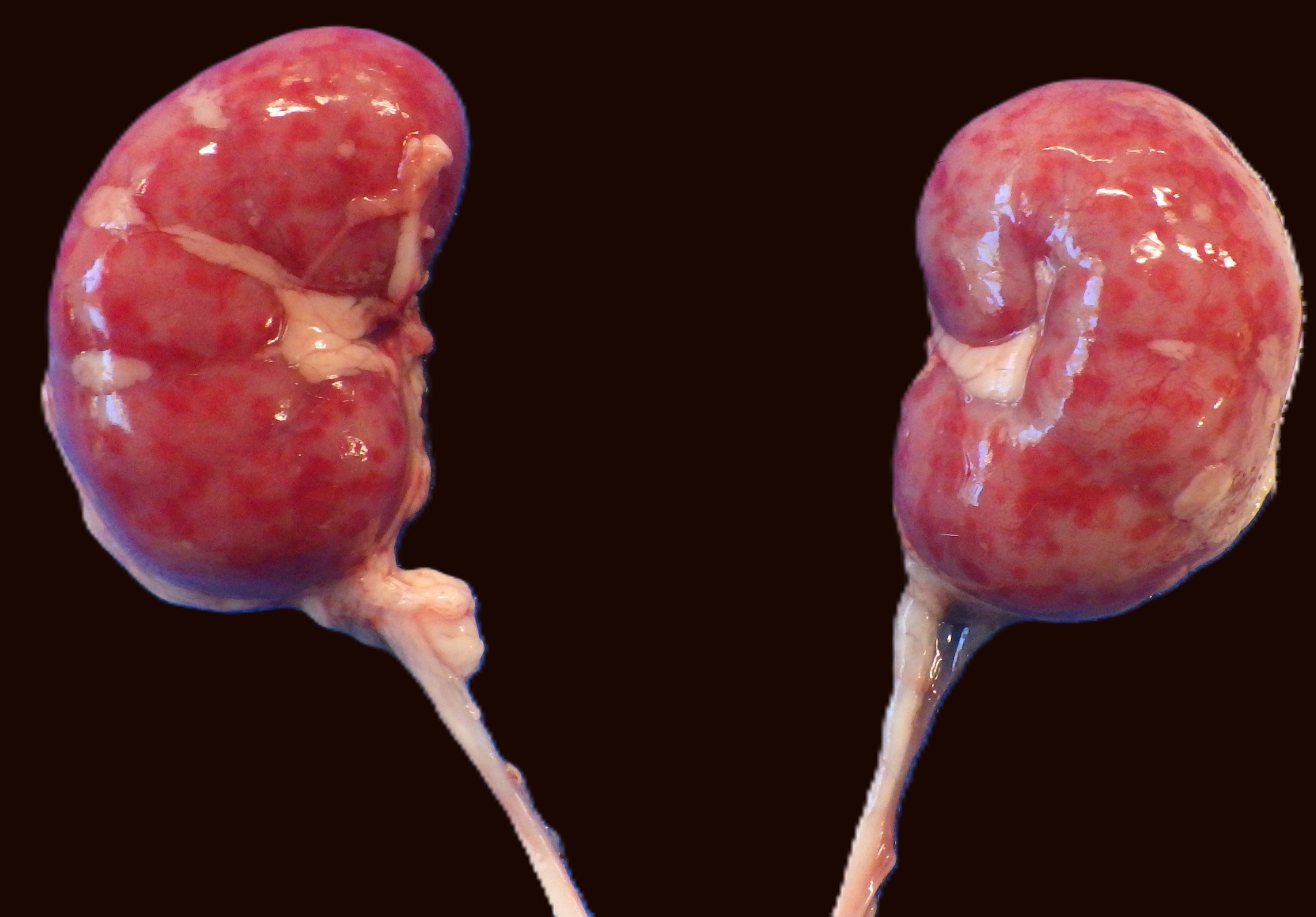
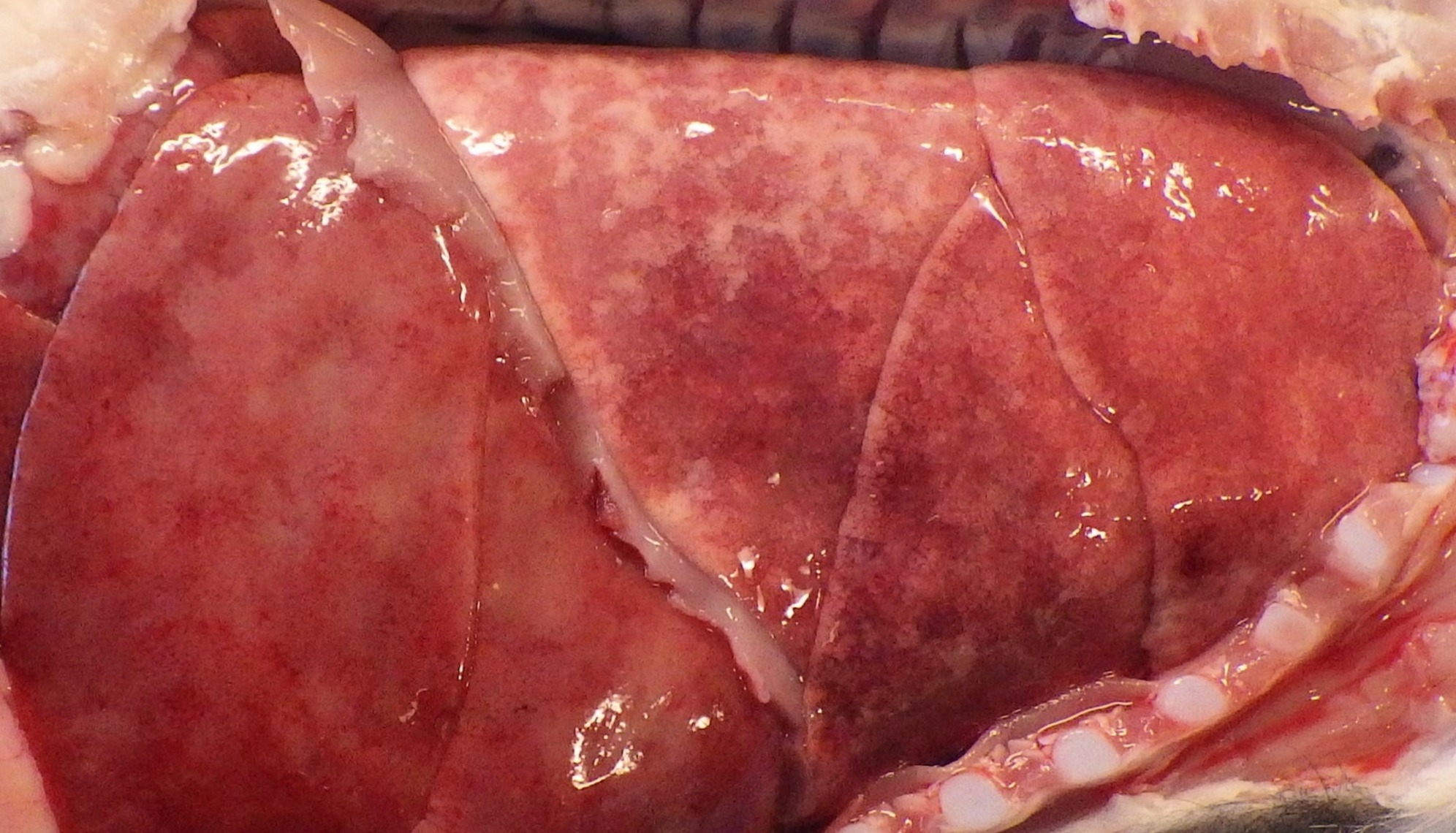
Gross Pathology: The lungs were mottled dark to light red, non-collapsing, rubbery, and wet on cut section. The right middle lung lobe sank in formalin. The liver contained multifocal, pinpoint (<1.0 mm) pale red to tan foci scattered throughout all lobes. Bilaterally, both kidneys had multifocal to coalescing hemorrhagic foci (0.1-0.2 cm in diameter) within the cortex, which extended into the corticomedullary junction on cut section.
Microscopic Description:
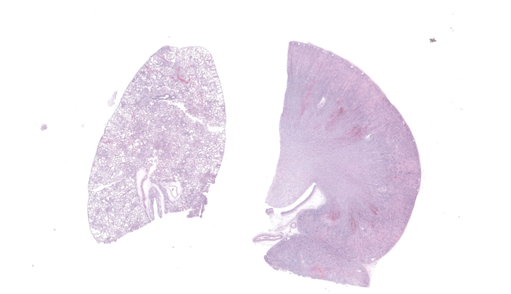
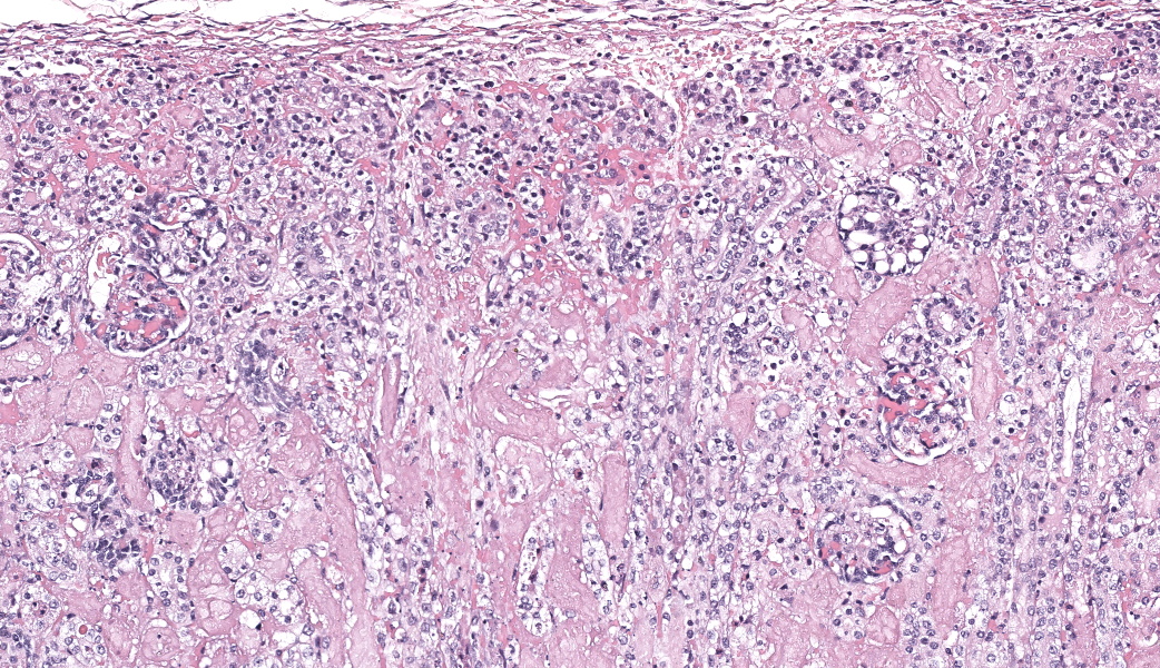
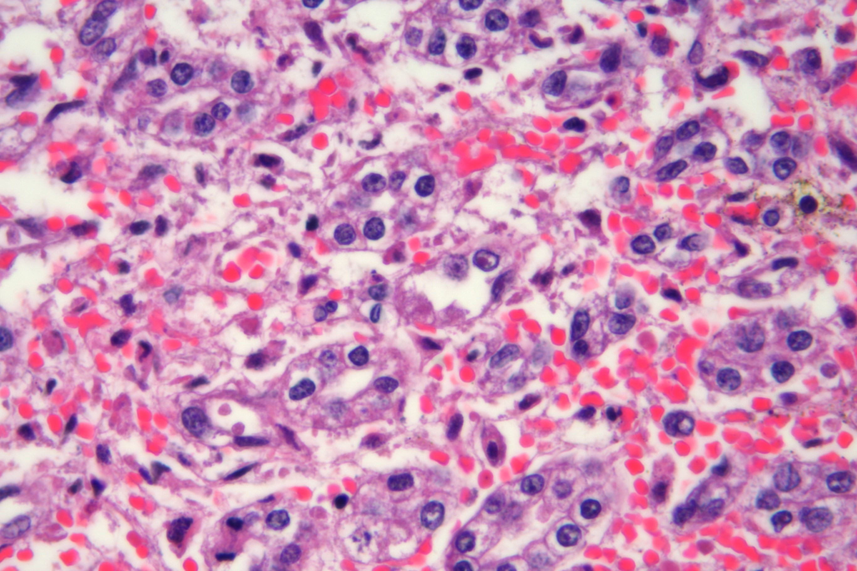
Kidney: Multifocally throughout the section, involving approximately 40% of the parenchyma, primarily within the cortex and extending to the corticomedullary junction, there are variably sized areas of necrosis. Affected tubules and occasional glomeruli are pale and eosinophilic, with loss of differential staining and absent to karyorrhectic nuclei, accompanied by interstitial hemorrhage and edema. Rarely, individual epithelial cell nuclei contain a single, round, eosinophilic intranuclear inclusion body that marginates the chromatin.
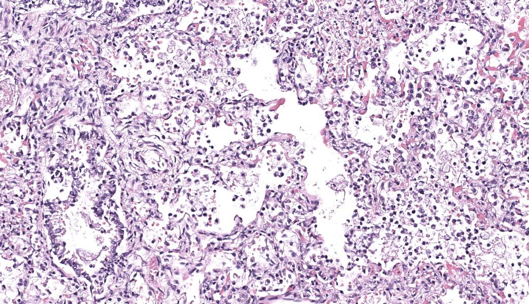
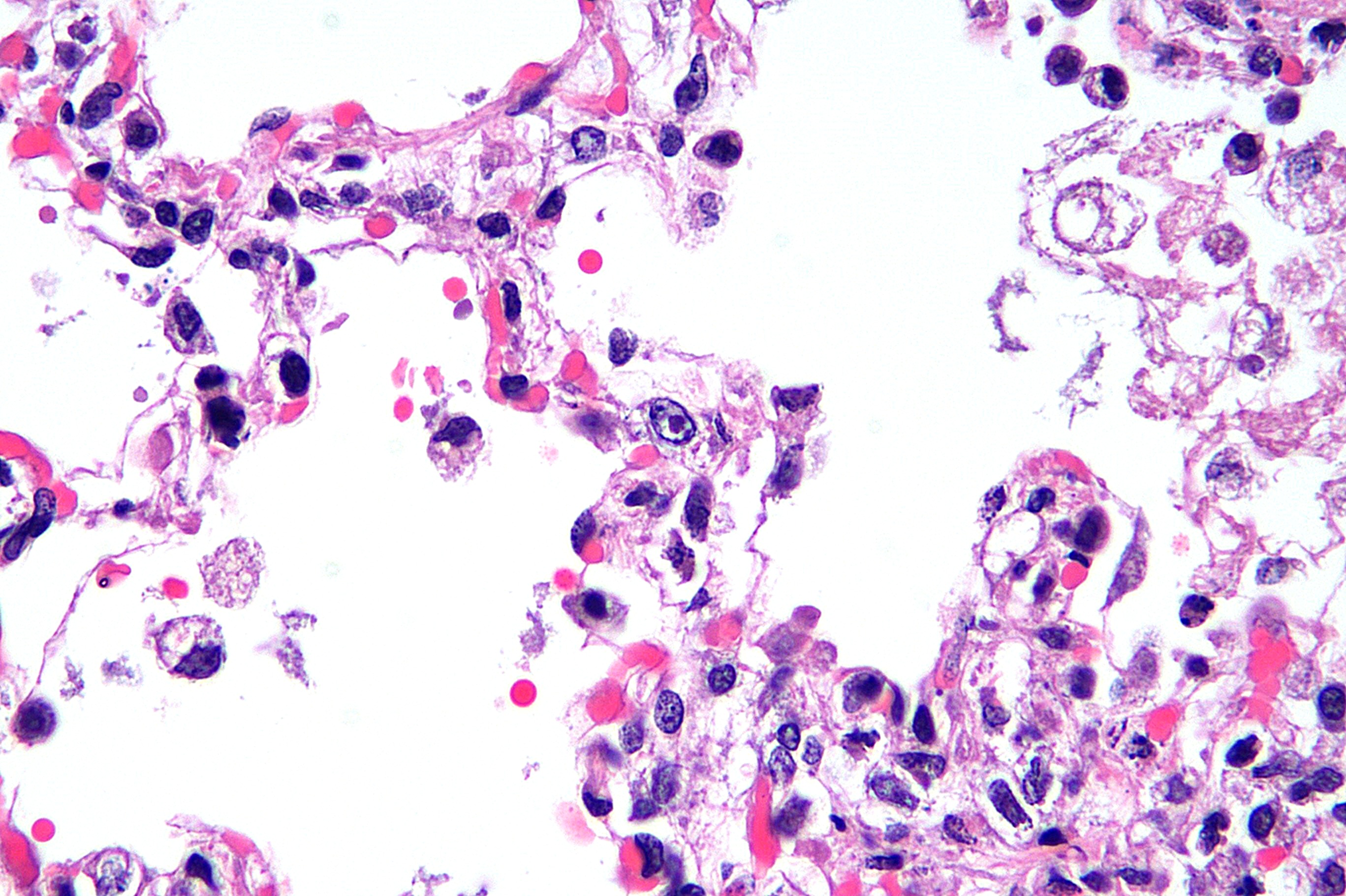
Lung: Multifocally throughout the section, alveolar septa are frequently and variably expanded, up to fourfold, by abundant fibrin, necrotic cellular debris, and very low numbers of neutrophils. Adjacent alveolar spaces contain similar material, along with small amounts of hemorrhage and foamy macrophages. Rarely, small eosinophilic intranuclear inclusion bodies are present within macrophages and pneumocytes at the margins of necrotic areas. The interstitium surrounding bronchi and the tunica adventitia of arteries is often expanded by a moderate amount of clear space (edema).
Contributor's Morphologic Diagnosis: Kidney, liver, brain, and lung: Nephritis, hepatitis, encephalitis, and interstitial pneumonia, necrotizing, multifocal, marked, with rare eosinophilic intranuclear inclusion bodies.
Contributor's Comment:
Canine herpesvirus type 1 (CHV-1) is an enveloped, double-stranded DNA virus in the Alphaherpesvirinae subfamily of the Herpesviridae family. First identified in the 1960s, it remains a significant cause of perinatal mortality in dogs.2,4 The virus replicates most efficiently at subnormal body temperatures (34-36°C), which helps explain why neonatal puppies, who cannot yet regulate their body temperature well, are particularly susceptible. CHV-1 is inactivated at temperatures above 40°C, underscoring the importance of external heat support in managing suspected cases.4
The clinical course of infection is strongly age-dependent. Typically, puppies less than three weeks of age are at risk, often developing a fulminant, systemic infection characterized by viremia, multisystemic necrosis, and high mortality. In contrast, older puppies and adult dogs typically develop localized infections affecting the upper respiratory tract, eyes, or genital mucosa.3,4 Infection during pregnancy in a naive bitch can result in fetal loss, abortion, or neonatal death. Surviving dogs may become latent carriers, harboring the virus in sensory ganglia with potential for reactivation during periods of stress, illness, or immunosuppression.2,3
Transmission in neonates occurs in utero, during passage through the birth canal, or postnatally via oronasal secretions from the dam or littermates. Fomites and caretakers may also contribute. In older dogs, aerosol transmission is more common. After local replication in the nasal mucosa and tonsils, the virus spreads to regional lymph nodes and subsequently disseminates to visceral organs and the central nervous system via infected leukocytes.2,4
The incubation period ranges from 6 to 10 days, and affected litters may experience mortality rates approaching 100% within a week of onset. Clinical signs include crying, anorexia, abdominal pain, hypothermia, and ataxia; some puppies may die suddenly with minimal premonitory signs.3
At necropsy, affected neonates often show characteristic gross and microscopic lesions, including multifocal hemorrhage and pallor in the liver, kidneys, lungs, spleen, and gastrointestinal tract, typically with minimal inflammation. Enlarged lymph nodes and nonsuppurative meningoencephalitis are also common. These findings are considered highly suggestive of systemic CHV-1 infection in puppies less than three weeks old.3,4
Contributing Institution:
Midwestern University College of Veterinary Medicine Diagnostic Pathology Center 5725 West Utopia Rd. Glendale, AZ 85308 https://www.mwuanimalhealth.com/diagnostic-pathology-center
JPC Morphologic Diagnosis:
- Kidney: Nephritis, necrotizing, acute, multifocal, moderate, with rare intranuclear viral inclusions.
- Lung: Pneumonia, necrotizing, acute, multifocal to coalescing, moderate, with rare intranuclear viral inclusions.
JPC Comment:
What a classic case! The contributor provides a thorough, well-written comment on this entity that has been seen several times throughout the years of the WSC due to its continued relevance. Dr. Carvallo made a point to mention how pathologists should use all the clues at their disposal to help them gather as much information as possible. In this case, the size of the organs was sufficiently small that one could assume they were from a very young animal. Most would diagnose the changes in the lung as consistent with an interstitial pneumonia, but Dr. Carvallo stated that the preferred term in current veterinary literature would be "interstitial lung disease." This term is currently being added into the pathology lexicon as a broad term for cellular damage +/- leukocyte infiltration that mainly targets the interstitium, including the alveolar septa and adjacent bronchioles.1 The term "interstitial pneumonia" is now being classified as a subtype of interstitial lung disease characterized by an increased number of leukocytes thickening the alveolar septa and vascular adventitia as seen with such diseases as ovine progressive pneumonia (OPP), porcine reproductive and respiratory syndrome (PRRS), sepsis, or parasitic larval migrations.1
References:
- Carvallo-Chaigneau FR, Caswell JL. Special section on interstitial lung disease in domestic animals. Journal of Veterinary Diagnostic Investigation. 2026;0(0).
- Decaro N, Martella V, Buonavoglia C. Canine adenoviruses and herpesvirus. Vet Clin North Am Small Anim Pract. 2008;38(4):799-814. doi:10.1016/j.cvsm.2008.02.006.
- Evermann JF, Ledbetter EC, Maes RK. Canine reproductive, respiratory, and ocular diseases due to canine herpesvirus. Vet Clin North Am Small Anim Pract. 2011;41(6):1097-1110. doi:10.1016/j.cvsm.2011.08.007.
- Greene CE. Canine herpesvirus infection. In: Greene CE, ed. Infectious Diseases of the Dog and Cat. 4th ed. Philadelphia, PA: WB Saunders; 2012:48-54