Signalment:
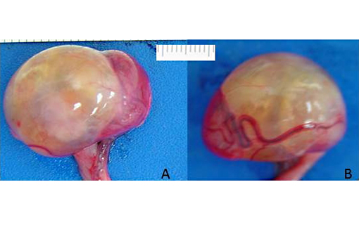
Gross Description:
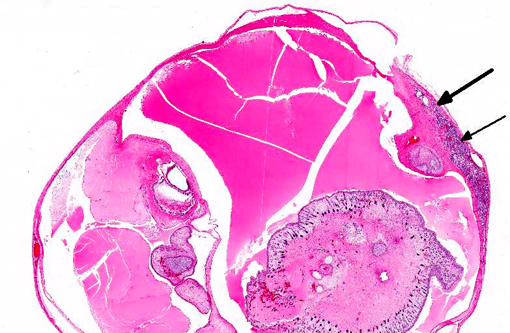
Histopathologic Description:
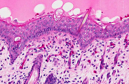
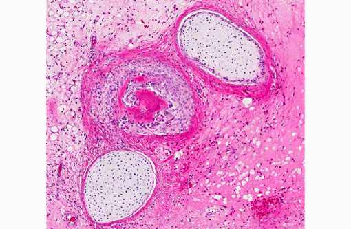
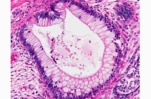
Ectodermal components include scattered nests of squamous epithelium with peripheral basal layers, central stratum lucidum, and inner stratum corneum and a central keratin core. Some sections contain haired skin with keratinized epidermis, hair follicles and shafts, dermal vasculature and fat, and clusters of adnexal glands. Mesodermal components include scattered islands of cartilage, rare spicules of new bone formation, and rudimentary tooth formation. Endodermal components consist of cystic spaces lined by complex cuboidal to columnar epithelium mixed with goblet cells that cover papillary villous-like projections and has a submucosa with lymphoid aggregates.
Morphologic Diagnosis:
Condition:
Contributor Comment:
Teratomas of the human testis can be classified as solid or cystic, and mature or immature, based on whether components have adult or embryonic features; the tumors often have features of both.(7) In animals, teratomas are most frequently found in the horse, the majority in cryptorchid testes.(3) In the fetus, teratoma formation probably prevents normal descent.(12) Human cases with undescended testes are 5-48 times more likely to be neoplastic(12), but neoplasia after surgical reduction (orchiopexy) and in the other normally descended testis is also reported.(1)
In animals most teratomas are benign, as are most ovarian teratomas in women, whereas most post pubertal testicular tumors in men are malignant (except those occurring in childhood), suggesting origin from benign and malignant cells respectively.(6) The difference may be based on the human females tolerance for parthenogenetic development of immature somatic ova cells into three germ layers while suppressing neoplastic cells, in contrast to the human male that differentiates malignant immature somatic cells less efficiently in the embryo.(11) Why this hypothesis does not appear to extend to animals is unexplained.
JPC Diagnosis:
Conference Comment:
For comparison to the testicular teratoma, conference participants examined a mouse ovarian teratoma, provided by the moderator. The ovarian teratoma was composed of haphazard regions of neuroectoderm, vague glandular/ductular components, poorly differentiated muscle (mesoderm) and respiratory epithelium (endoderm), consistent with an immature teratoma.Â
Table 1: Embryonic germ cell layers and selected tissue derivatives.(2,10)
| Ectoderm | Mesoderm | Endoderm |
|
|
|
References:
2. Foster RA. Male reproductive system. In: McGavin MD, Zachary JF, eds. Pathologic Basis of Veterinary Disease. 5th ed. St. Louis, MO: Elsevier; 2012:1142-1143.
3. Foster RA, Ladd PW. Male genital system. In: Maxie MG, ed. Jubb, Kennedy, and Palmers Pathology of Domestic Animals. 5th ed. Vol. 3. St. Louis, MO: Elsevier Limited; 2007:565-619.Â
4. Jamadagni SB, Jamadagni PS, Lacy SH, et al. Spontaneous nonmetastatic choriocarcinoma, yolk sac carcinoma, embryonal carcinoma, and teratoma in the testes of a Swiss albino mouse. Toxicol Pathol. 2013; 41(3):532-536.
5. Keller DL, Schneider LK, Chamberlin T, Ellison M, Steinberg H. Intramedullary teratoma in a domestic ferret. J Vet Diagn Invest. 2012;24(3):621-624.Â
6. Lakhoo, K. Neonatal teratomas. Early Hum Develop. 2010;86:643-647.
7. Moulton JE. Tumors of the genital system. In Tumors of Domestic Animals. 2nd ed. Berkely, CA: University of California Press; 1978:309-345.Â
8. Murai A, Yanai T, Kato M, Yonemaru K, Sakai H, Masegi T. Teratoma of the umbilical cord in a giraffe (Giraffa camelopardalis reticulata). Vet Pathol. 2007;44(2):204-206.
9. Robinson NA, Manivel JC, Olson EJ. Ovarian mixed germ cell tumor with yolk sac and teratomatous components in a dog. J Vet Diagn Invest. 2013;25(3):447-452.
10. Schlafer DH, Miller RB. Female genital system. In: Maxie MG, ed. Jubb, Kennedy, and Palmers Pathology of Domestic Animals. 5th ed. Vol. 2. Philadelphia, PA: Saunders Elsevier; 2007:450, 453-4.Â
11. Ulbright TM. Germ cell tumors of the gonads: a selective review emphasizing problems in differential diagnosis, newly appreciated, and controversial issues. Modern Path. 2005;18:S61-S79.
12. Yam B, Georgiou NA, Khullar P, et al. Radiology-Pathology conference: mature teratoma arising from an intra-abdominal undescended testis in a 7-month-old infant. Clin Imaging. 2010;34:466-471.