Signalment:
Gross Description:
Morphologic Diagnosis:
Condition:
Contributor Comment:
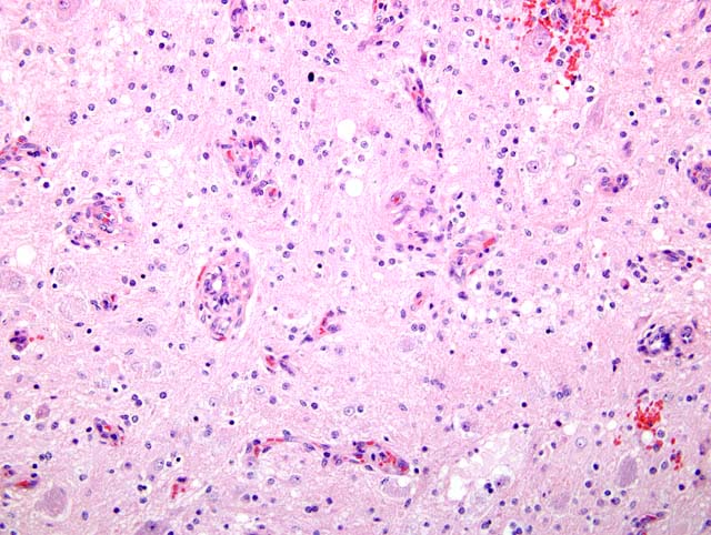
Diagnosis can be confirmed by a variety of methods to include transmission electronmicroscopy (TEM), histochemistry, and immunohistohemistry. TEM reveals dense core vesicles in ganglion cells and neuronal processes. Umyelinated axons are encased by Schwann cells. Histochemical stains for axons and myelin reveal that nerve fibers are nonmyelinated. Immunohistochemical stains revel that ganglion cells are positive for NSE and Schwann cells are positive for S-100 and vimentin. Nerve fibers are positive for neurofilament protein.(4)
Neurogenic tumors of the canine ovary have not been described. The nerve supply to the ovary is via a sympathetic plexus that accompanies the ovarian vessels. This relationship may explain the numerous small vessels enmeshed in the tumor.
JPC Diagnosis:
Conference Comment:
Although teratomas are usually defined as neoplasms composed of tissue derived from at least two germinal layers (6), occasional ovarian tumors may be composed of cells from only one layer. In humans, these tumors are derived predominately or exclusively of endodermal or ectodermal tissue and are referred to as monodermal teratomas.(7) Rarely these neoplasms are composed almost exclusively of neuroectodermal tissue, including astrocytes, oligodendroglial cells, and ganglion cells.(7) Arguably such tumors may be hamartomas rather than true neoplasms.Â
References:
2. Cole DE, Migaki,G, Leipold HW: Colonic ganglioneuromatosis in a steer. Vet Pathol 27:461-462, 1990
3. Fairley, McEntee MF: Colorectal ganglioneuromatosis in a young female dog (Lhasa Apso). Vet Pathol 27:206-207, 1990
4. Ribas JL, Kwapien RP, Pope ER: Immunohistochemistry and ultrastructure of intestinal ganglioneuroma in a dog. Vet Pathol 27:376-379, 1990
5. Schlafer DH, Miller RB: Female genital system. In: Jubb, Kennedy, and Palmer's Pathology of Domestic Animals, vol 3 ed. Maxie MG, pp. 450-456. Elsevier Limited, Philadelphia, PA, 2007
6. MacLachlan NJ, Kennedy PC: Tumors of the genital systems. In: Tumors in Domestic Animals, ed. DJ Meuten, 4th ed., p. 554. Blackwell, Ames, IA, 2002
7. Scully RE, Young RH, Clement PB: Monodermal Teratomas. In: Atlas of Tumor Pathology, Tumors of the Ovary, Maldeveloped Gonads, Fallopian Tube, and Broad Ligament, ed. J Rosai, Third series, Fascicle 23, pp. 285-306, Armed Forces Institute of Pathology, Washington, DC, 1996