Signalment:
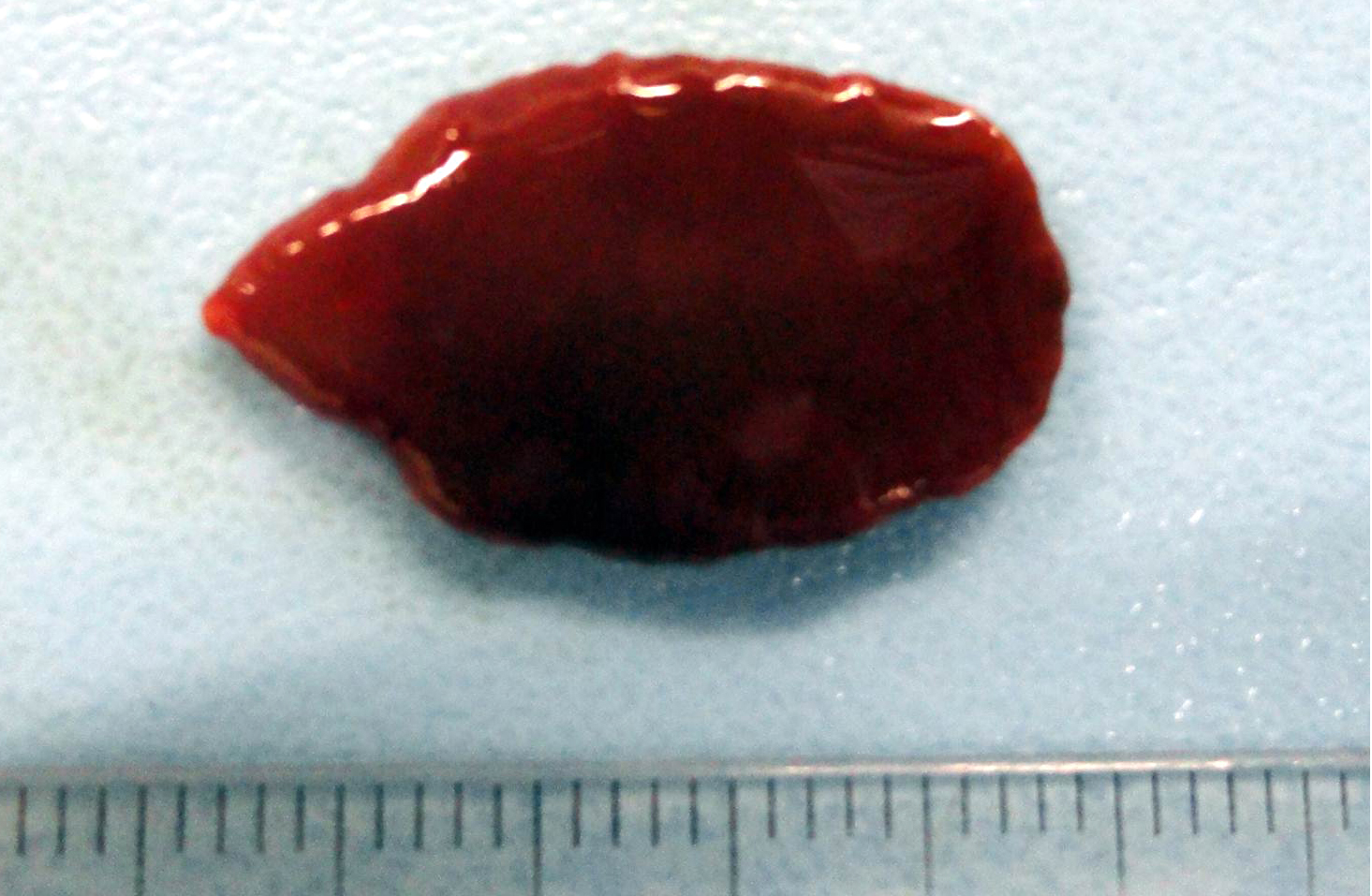
Gross Description:
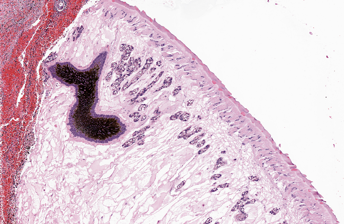
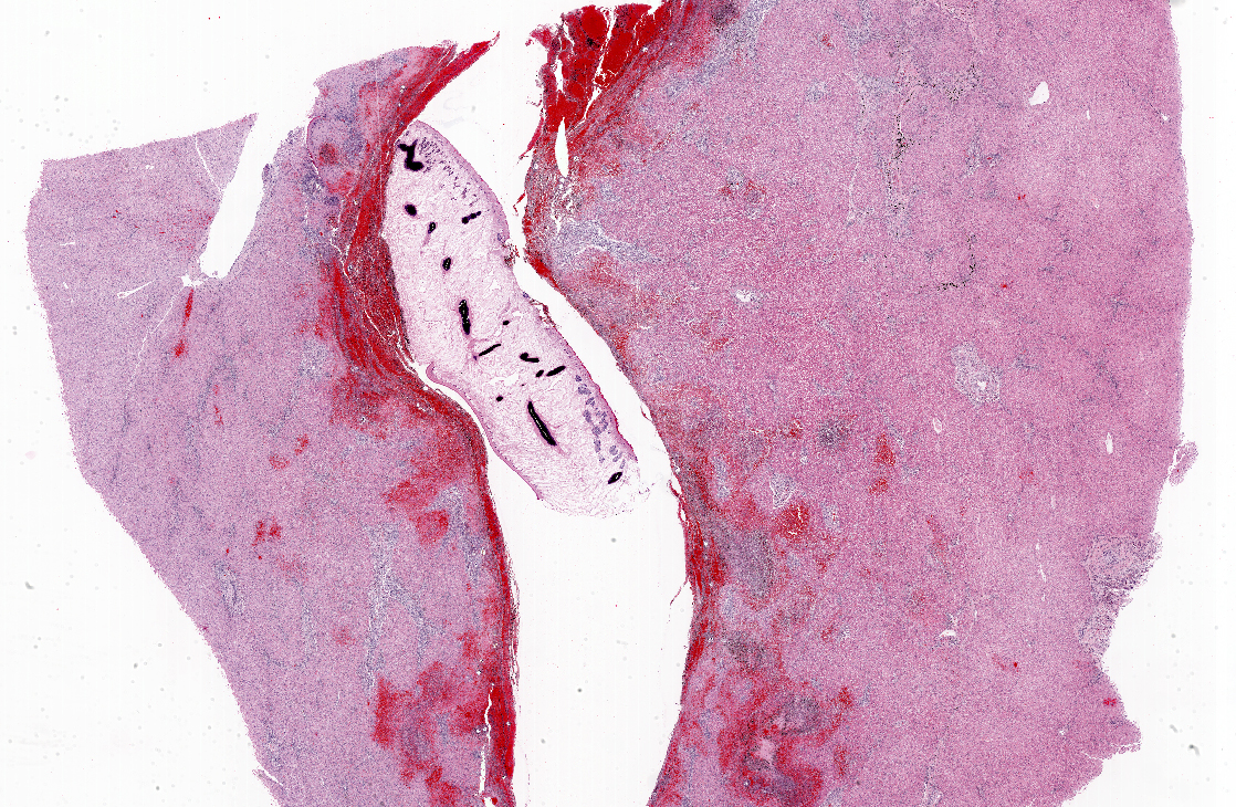
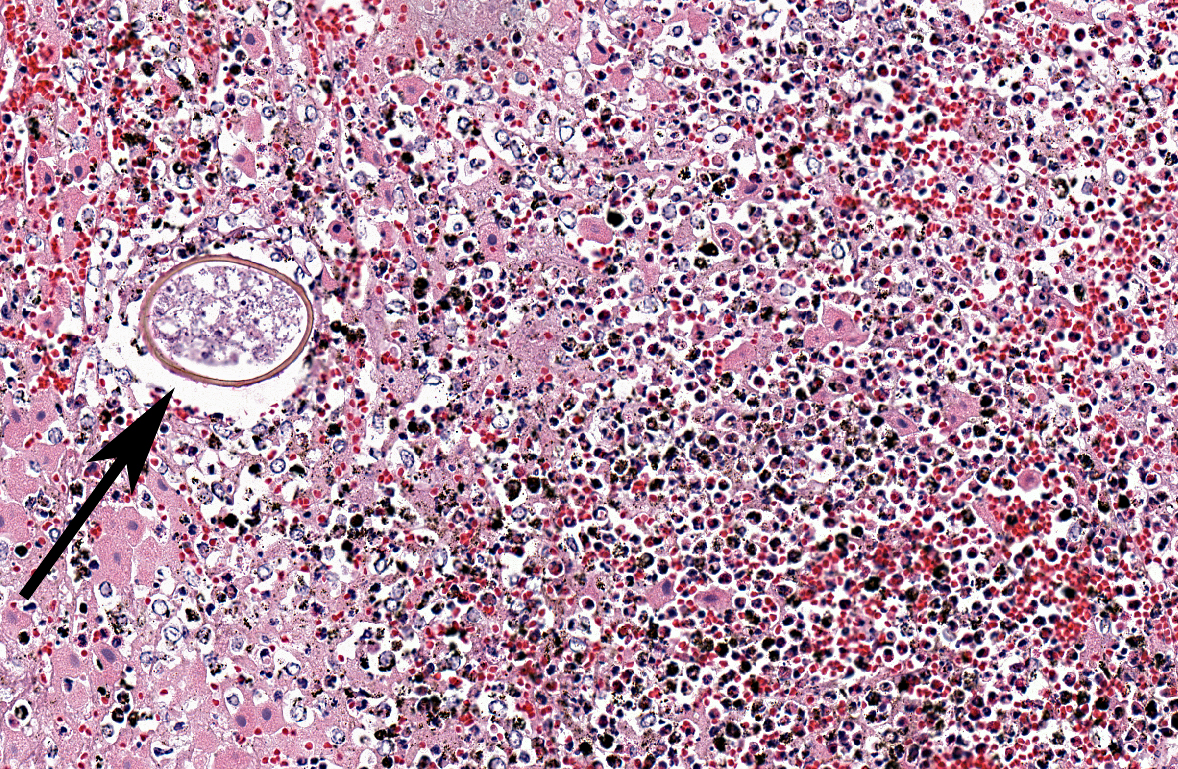
Histopathologic Description:
Acutely, hemorrhage and eosinophils predominate. Stranded in the fibrosis of chronic lesions or in hemorrhage in acute lesions are scattered operculate ova, each with a well defined yellow-brown shell and a central developing embryo. Some ova are degenerate, with neutrophils or multinucleate phagocytes occur around them. Adult trematodes are present in acute migratory tracts surrounded by hemorrhage, and are not located in bile ducts. They are characterized by an external tegument and absence of a body cavity. Muscular, external suckers are present in some sections. The body is filled by loose, pale eosinophilic parenchyma and suspended within is the intestinal tract, containing brown pigment like that seen in tissue. Vitelline bodies are also sometimes apparent. Both male are female reproductive organs can be seen in some sections. A mixture of eosinophils and lymphocytes is present in peripheral hemorrhage and in the adventitia of portal triads. In areas of acute migration, hepatocytes have undergone localized necrosis without reference of their position in the lobule. Most inflamed portal areas contain increased bile duct profiles.
Morphologic Diagnosis:
Lab Results:
Condition:
Contributor Comment:
JPC Diagnosis:
Conference Comment:
A primary differential in this case is Fasciola hepatica, which can be as large as F. magna and may appear similar histologically. In cattle, these two flukes can be differentiated, though, by the location of the adult flukes the liver and the presence of considerable iron porphyrin pigment in Fascioloides magna infections. Adults of F. hepatica are present in bile ducts, while F. magna adults are distributed throughout the liver parenchyma. The migration tracts produced by larval F. hepatica do not generally cause sufficient damage to alter liver function in the host, as may be the case with F. magna. However, clinically important disease can be caused by F. hepatica in cattle and sheep when migrating larvae produce areas of local coagulative necrosis and lower hepatic oxygen tension. The areas of hypoxia create a favorable environment for the germination of Clostridium noyvi spores, resulting in a form of necrotizing hepatitis known as black disease. C. noyvi releases an alpha toxin and the necrotizing and hemolytic beta toxin, lecithinase. In Western and Eastern Europe, black disease may be incited by Dicrocoelium dendriticum. Bacillary hemoglobinuria, caused be the closely related C. haemolyticum, may also occur secondary to F. hepatica migration and has a similar pathogenesis to that of black disease(4).
When Fascioloides magna encysts in the liver, the resulting 2-5 cm diameter encapsulated, pigmented cysts may grossly resemble melanocytic tumors.
References:
2 McClanahan SL, Stromberg BE, Hayden DW: Natural infection of a horse with Fascioloides magna. J Vet Diagn Invest 17:382-385,2005.
3 Novobilsky A,Horackova E, Hirtova L, et al: The giant liver fluke Fascioloides magna (Bassi 1875) in cervids in the Czech republic and potential of its spreading into Germany. Parasitol Res 100:549-553, 2007.
4 Stalker MJ, Hayes MA: Liver and biliary system. In Jubb, Kennedy and Palmers Pathology of Domestic Animals, ed. M.G. Maxie, 5th, ed., vol. 2, p354-362. Sanders Elsevier, St. Louis MO, 2007.