Signalment:
2-year-old, female Scottish Highland heifer,
Bos TaurusThis heifer had a history of mild respiratory difficulty since birth. In the last month, multiple cutaneous nodules developed on the head, thorax and hind limbs, with pus draining from a few. This animal was the only one affected on the farm.
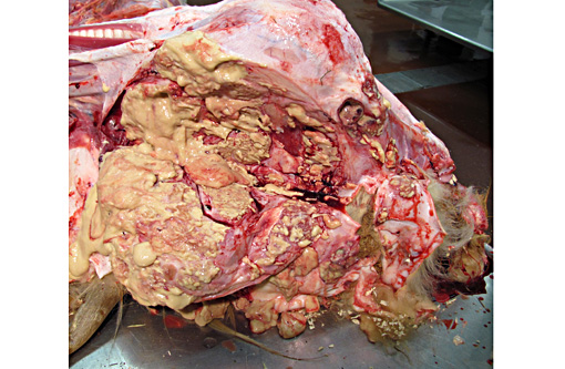
Gross Description:
The animal was in poor body condition. There was diffuse facial swelling and deformity. On cut section, there were numerous encapsulated and fistulating abscesses, up to 5 cm in diameter, in the subcutaneous tissues and muscles of the maxillary and mandibular regions, and, minimally, the tongue; the pus was thick, yellowish, and no "sulfur granules" were detected. Similar abscesses were also present in: 1) the walls of the pharynx and proximal esophagus, 2) the cervical, prescapular, inguinal and tracheobronchial lymph nodes, 3) the subcutaneous tissues of the thorax and both hind limbs, and 4) the lungs. Multiple, often coalescing ulcers, 4 mm in diameter, were observed on the gingiva, tongue, soft palate and, to a lesser degree, esophagus.
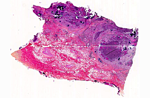
Histopathologic Description:
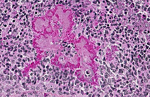
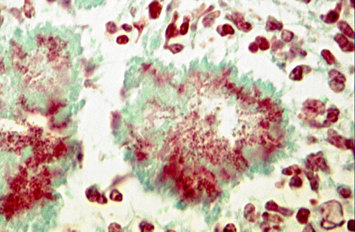
The submitted slide is a section of pharyngeal/cranial esophageal wall stained with hematoxylin-eosin-phloxin-saffron. The normal architecture is extensively obliterated by irregularly sized collections of degenerate neutrophils with variable numbers of surrounding and/or admixed macrophages (pyogranulomas), and dense fibrovascular (granulation) tissue infiltrated mainly by lymphocytes and plasma cells between pyogranulomas. Within these pyogranulomas, there are numerous structures composed of pale amphophilic, finely granular material (bacteria) surrounded by radiating, deeply eosinophilic, club-shaped material (Splendore-Hoeppli phenomenon). This material is multifocally and variably mineralized, with occasional associated multinucleated giant cells. A Gram stain showed the bacteria to be Gram-negative coccobacilli.Â
Morphologic Diagnosis:
Severe, multifocal chronic pyogranulomatous pharyngitis/esophagitis with Splendore-Hoppli material and intralesional Gram-negative coccobacilli.
Lab Results:
The heifer tested negative for bovine viral diarrhea pestivirus by fluorescent antibody testing (FAT) on the oral and esophageal mucosa. Bacterial culture of abscesses in skin, lymph nodes and lung yielded a heavy growth of
Actinobacillus lignieresii, in pure culture except in one lymph node in which
Trueperella pyogenes was also identified.
Condition:
Actinobacillus lignieresi
Contributor Comment:
The final diagnosis was actinobacillosis. Actinobacillosis is mainly a disease of soft tissues of cattle and sheep, but it has also been reported in other species including pigs, goats, horses and dogs.Â
Actinobacillus lignieresii is an opportunistic Gram-negative coccobacillary bacterium that is part of the normal flora of the oral cavity and rumen of cattle and sheep. Following trauma to the oral mucosa (e.g. plant material, teeth, etc...) or skin,
A. lignieresii invades the underlying soft tissues and typically causes pyogranulomatous lesions, often with lymphagenous extension to regional lymph nodes (and thus lymphangitis and lymphadenitis).(1,2,3) Iatrogenic cases have been described, e.g. surgical wound contamination during cesarean sections.(1,3)
Actinobacillus lignieresii is also a cause of bite wound infections in humans, especially horse bites.(3,4) Grossly, the lesions appear as variably-sized abscesses with abundant fibrous tissue, in which small granules, known as "sulfur granules", are usually seen. Microscopically, these lesions correspond to pyogranulomas, with abundant inflamed fibrous/fibrovascular tissue, centered on masses of coccobacilli surrounded by radiating club-shaped eosinophilic material (Splendore-Hoeppli phenomenon), and grossly seen as the granules.(2,3) Splendore-Hoeppli material can be seen in some bacterial, fungal and parasitic infections, but also in some non-infectious conditions; it is considered to be made up of immune complexes,(2) among others. Other bacteria that can cause similar microscopic lesions in cattle include
Staphylococcus aureus and
Pseudomonas aeruginosa (botryomycosis), and also
Actinomyces bovis; the latter causes mainly a mandibular osteomyelitis, known as "lumpy jaw", but can sometimes involve soft tissues.(1,2,7)
In cattle, the most frequent presentation is a chronic glossitis known as "wooden tongue", due to the very firm consistency imparted by the abundant fibrous tissue.(1,2) The disease is usually sporadic, but small outbreaks have been reported.(1,2,3) Other reported locations include the skin of the head, neck and limbs, forestomachs, lungs, pharynx, esophagus and lungs.(1,2,3) Clinical signs depend on lesion size and distribution; subclinical lesions, especially in the tongue and lymph nodes of the head and neck, may be found as incidental findings at slaughter.(3) Intravenous sodium iodine is the treatment of choice, but successful surgical exeresis of localized lesions has been described.(1,7) In the present case, the tongue was minimally involved and no sulfur granules were observed; an underlying cause was not found. Dystrophic mineralization was prominent, especially in "older" lesions. The multiple ulcerations in the oral and proximal esophageal mucosae were considered to be the result of
A. lignieresii infection, as they were associated with underlying pyogranulomatous lesions.Â
JPC Diagnosis:
Fibrovascular tissue: Cellulitis, pyogranulomatous, diffuse, severe, with Splendore-Hoeppli phenomenon, and numerous bacterial colonies
Conference Comment:
This is a classic case with challenging tissue identification as most sections lack discernible anatomic landmarks; however, the Splendore-Hoeppli phenomenon and club colonies leave few differentials for this lesion in the ox. Wooden tongue may be most aptly confused with lumpy jaw, the other classic large colony forming bacterial entity of the bovine oral cavity. Both
Actinobacillus lignieresii and
Actinomyces bovis form club colonies, but the colonies in actinomycosis are much larger with smaller and less discrete clubs.(6) The two can be readily distinguished by the invasion of bone in lumpy jaw or by gram stain as only
Actinomyces is gram positive.(2)
As the contributor nicely explains, the bacteria is considered normal flora thus most cases of wooden tongue are sporadic with prevalence at slaughter of up to 3.6%. Interestingly, herd outbreaks have occurred with up to 73% morbidity and are likely associated with abrasive feedstuffs and crowded conditions.(4)
References:
1. Angelo P, Alessandro S, Noemi R, et al. An atypical case of respiratory actinobacillosis in a cow. J Vet Sci. 2009 Sep;10(3):265-7.
2. Brown CC, Baker DC, Barker IK. Alimentary system. In: Maxie MG, ed. Jubb, Kennedy, and Palmers Pathology of Domestic Animals, Vol. 2, 5th ed., pp. 1-296. Elsevier Saunders, Philadelphia, PA, 2007.
3. Cahalan SD, Sheridan L, Akers CR, et al. Atypical cutaneous actinobacillosis in young beef cattle. Vet Rec. 2012 Oct 13;171(15):375.
4. Jones SL, Smith BP. Diseases of the alimentary tract. In: Smith BP, ed. Large Animal Internal Medicine. 4th ed. St. Louis, MO: Mosby Elsevier; 2009:782-784.
5. Peel MM, Hornidge KA, Luppino M, et al. Actinobacillus spp. and related bacteria in infected wounds of humans bitten by horses and sheep. J Clin Microbiol. 1991 Nov;29(11):2535-8.
6. Thompson K. Bones and joints. In: Maxie MG, ed. Jubb, Kennedy, and Palmers Pathology of Domestic Animals, Vol. 2, 5th ed. Philadelphia, PA: Elsevier Saunders; 2007:98-99.
7. Thompson PN, Van der Lugt JJ, Olivier-Carstens A. Botryomycosis associated with Pseudomonas aeruginosa in the nasopharynx of a cow. Vet Rec. 2001 Oct 20; 149(16):495-6.