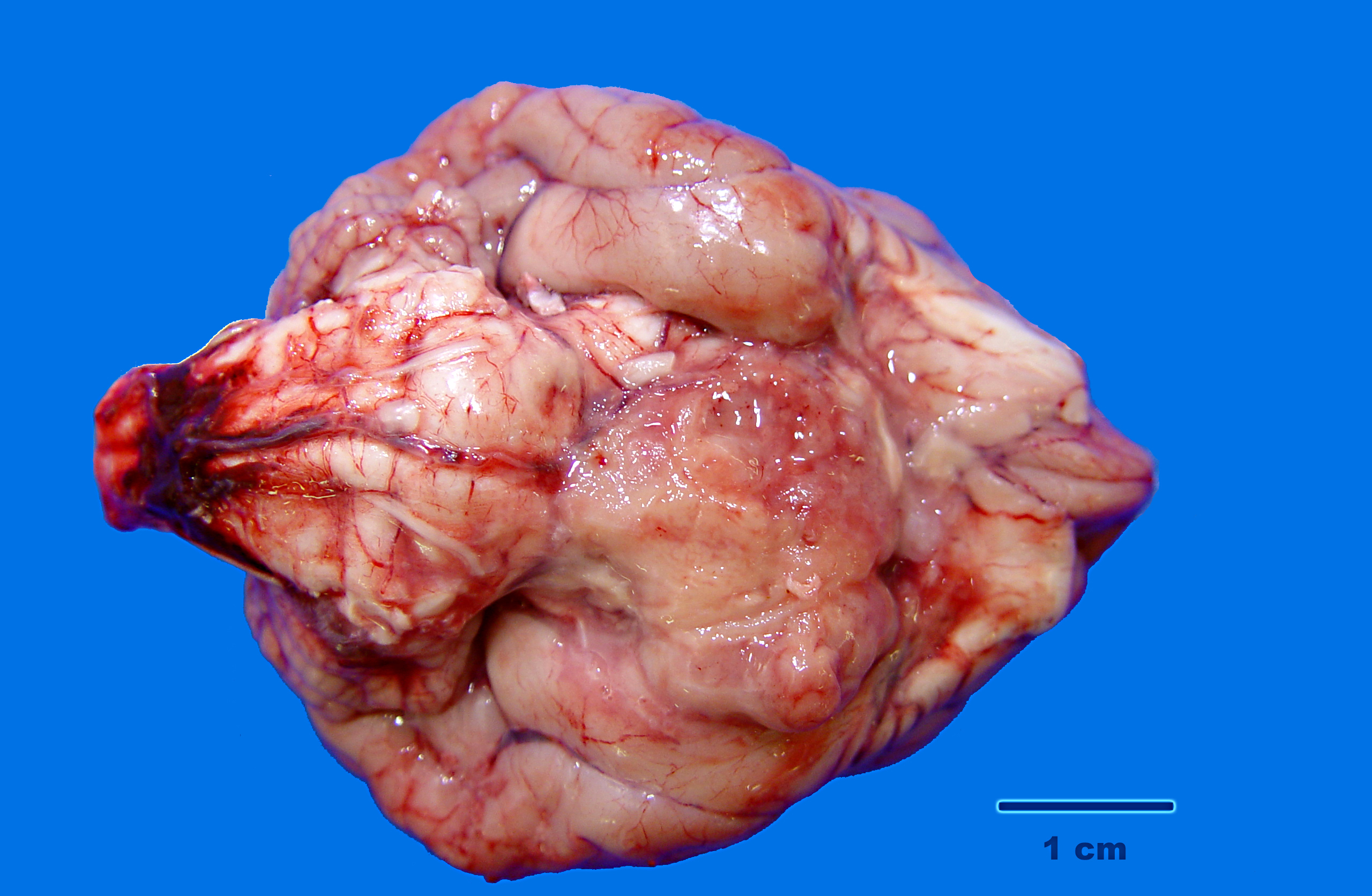
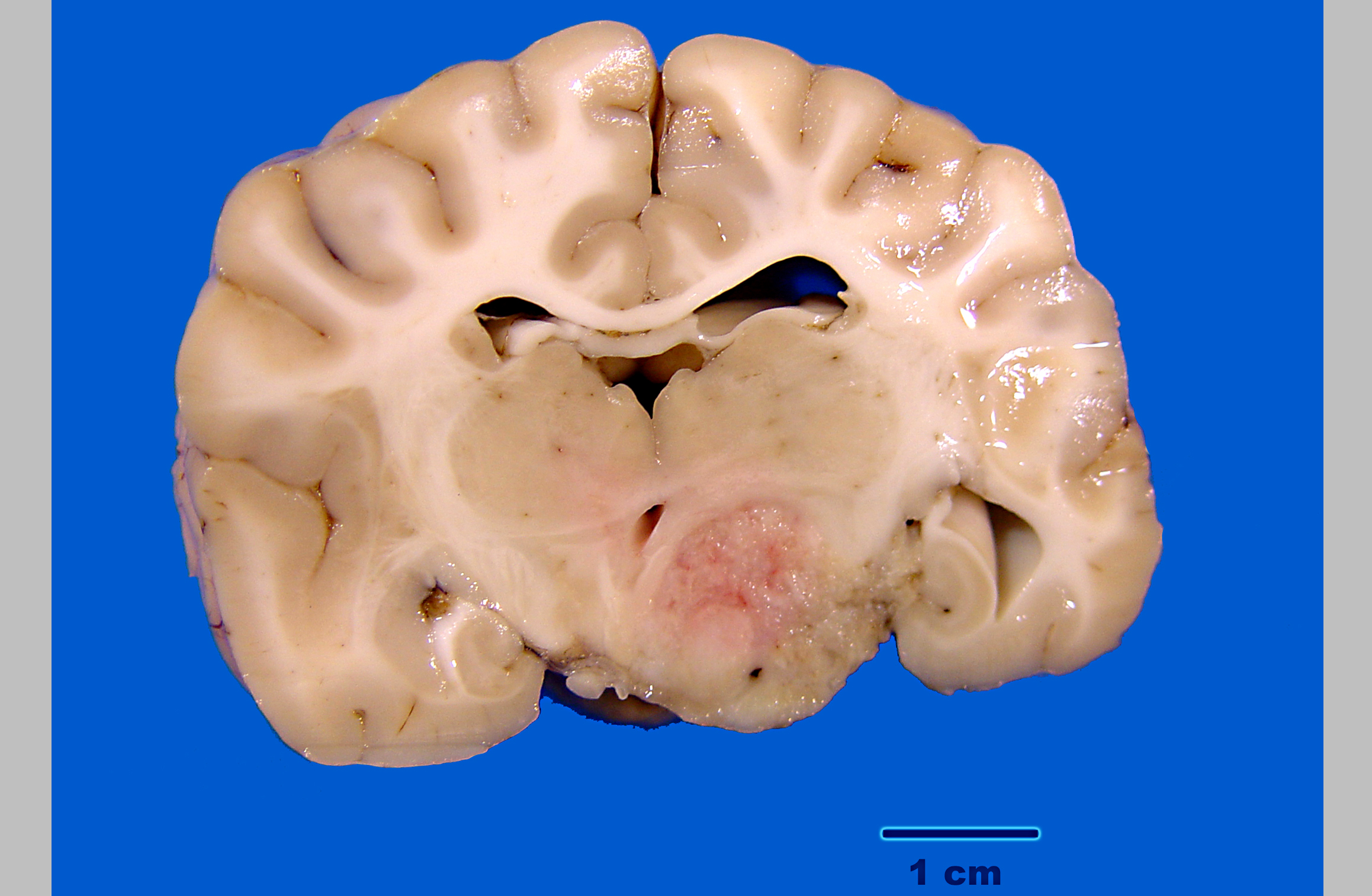
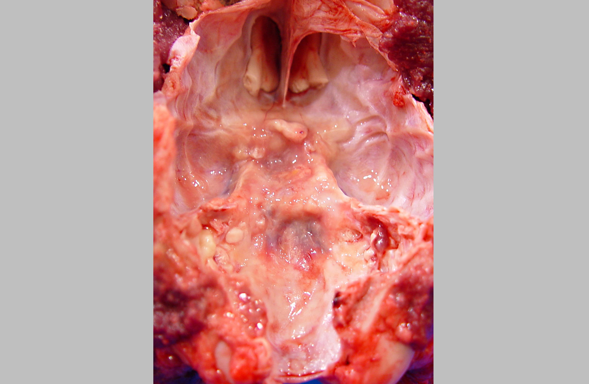
Signalment:  

5-year-old, male/castrated, American Pit Bull Terrier, dog, Canis familiars.The dog had a two week history of listlessness and decreased appetite. When examined at the clinic, the animal was dehydrated, lethargic, showing neurological signs (standing in the corner staring at the wall) and had anisocoria. Pain was elicited on flexion and extension of the neck. An intracranial mass was suspected and the owners opted to euthanize the dog.


Gross Description:  

The carcass is in good body condition; significant changes are restricted to the brain/skull base. A soft, pink, irregular mass measuring approximately 1 x 2 x 2 cm is present on the ventral surface of the brain, centered over the diencephalon slightly to the left of midline. The mass extends caudally from the olfactory tract to the rostral margin of the pons. Laterally the mass abuts the piriform lobe on the right and covers half of the piriform lobe on the left. On cut section, the mass infiltrates and compresses the hypothalamus, thalamus, piriform lobe and the hippocampus. The underlying floor of the skull appears irregular, thickened, and slightly yellow; the pituitary gland could not be identified.


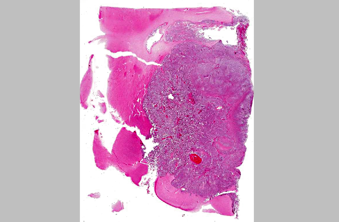
Histopathologic Description:

A well-delineated, nonencapsulated, moderately cellular, multilobulated mass compresses and invades the neuropil of the thalamus, hypothalamus, hippocampus, piriform lobe, optic tract, and the pituitary gland (depending on the section). Three cell types comprise the mass. Nests and solid lobules of loosely packed neoplastic cells separated by fine collagenous septa predominate; these cells are round to polygonal with distinct cell margins, a scant to moderate amount of flocculent amphophilic cytoplasm and a central round nucleus with granular to coarsely clumped, often hyperchromatic, chromatin and an indistinct nucleolus. Scattered amongst these cells are discrete islands and trabeculae of large polygonal cells with moderate amounts of eosinophilic cytoplasm and a large oval vesicular nucleus with a single distinct nucleolus; these cells often contain discrete cytoplasmic vacuoles which displace the nucleus to the periphery or, less often, exhibit squamous differentiation. Rare aggregates of tubule like structure lined by one to multiple layers of cuboidal to columnar epithelial cells with basal nuclei, granular chromatin and single to multiple nucleoli comprise the third population (not present in all slides). There is moderate anisocytosis and anisokaryosis. Mitoses, which are largely restricted to the round cells, average between 2 and 3 per HPF (40X). Small to moderate numbers of lymphocytes form multifocal aggregates within the supporting stroma, often surrounding blood vessels. Variably sized foci of necrosis and/or hemorrhage are scattered throughout the mass and rare mineral deposits and intravascular fibrin thrombi are noted.


Morphologic Diagnosis:  

Malignant mixed germ cell tumor, brain (Canine suprasellar germ cell tumor).


Lab Results:  

Paraffin embedded tissue sections were sent to the Diagnostic Center for Population and Animal Health (Michigan State University). Sections were evaluated for Alphafetoprotein immunohi s tochemical marker s . Approximately 60-70 % of the atypical cell population had strong positive cytoplasmic labeling with Alphafetoprotein.


Condition:  

Suprasellar germ cell tumor


Contributor Comment:  

Tumors in the seller region of the canine brain include pituitary tumors, craniopharyngioma, and suprasellar germ cell tumor.(8) Histological features in this case were not compatible with a pituitary tumor. Suprasellar germ cell tumors may be misclassified as craniopharyngioma, and this diagnosis was considered in this case.(6,14) A diagnostic feature of craniopharyngioma in humans is an epithelial component consisting of outer palisading columnar cells with central stellate cells resembling ameloblastoma; this characteristic was not observed in this mass.(10,14)

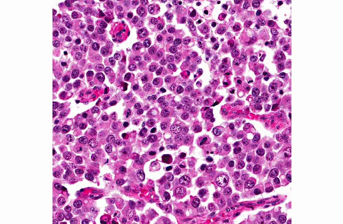
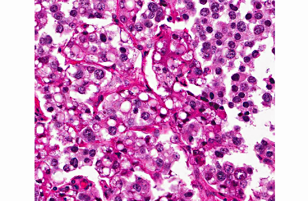
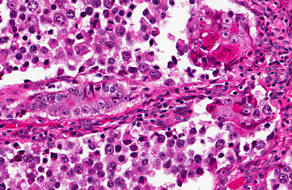
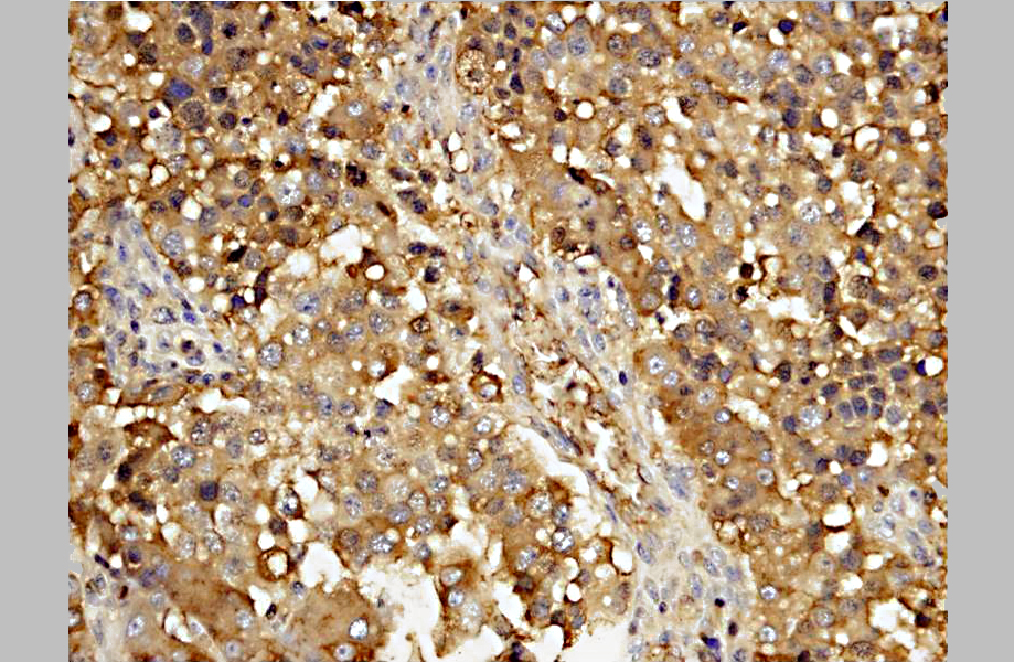
A diagnosis of suprasellar germ cell tumor was made based on three criteria: 1) midline suprasellar location, 2) presence within the tumor of several distinct cell types; one population resembling seminoma or dysgerminoma and others suggesting teratomatous differentiation into secretory glandular and squamous elements, and 3) positive staining for alphafetoprotein.(14) Alpha-fetoprotein is a commonly used marker for germ cells in human and has been shown to be a useful marker for canine intracranial germ cell tumors.(14)

Germ cell tumors most commonly occur in the ovaries and testes (dysgerminomas and seminomas respectively). Extragonadal germ cell tumors, with similar histology, are reported less commonly and typically occur on midline, affecting the mediastinum, brain, and coccyx in humans.(13) In dogs, these rare tumors have been reported in the brain, the eye, and the spinal cord.(5,10,11,12,14) Intracranial germ cell tumors tend to localize in the pineal and suprasellar regions in humans, while the suprasellar region is preferentially affected in dogs.(14) Suprasellar germ cell tumors have also been described in three lake whitefish (Coregonus clupeaformis).(9)

Presenting clinical signs in affected dogs include visual disturbance, pupil abnormality, lethargy, anorexia, and less often, signs of pituitary dysfunction.(10,12,14) Typically young adult dogs are affected and there is a possible breed predilection for the Doberman Pinscher.(14)

The pathogenesis of these tumors remains poorly understood, with the main question being the origin of the germ cells in the brain.(6) During embryonic development, progenitor germ cells originate in the epiblast. These germ cells then migrate to the caudal yolk sac endoderm and reach the gonadal ridge via the dorsal mesentery of the hindgut. Extragonadal germ cell tumors are presumed to arise from ectopic embryonic germ cells which may become displaced during development, survive physiologic dissolution, and undergo neoplastic transformation.(3,6)


JPC Diagnosis:  

Cerebrum: Suprasellar germ cell tumor.


Conference Comment:  

The contributor provided a very good summary of suprasellar germ cell tumors. In describing the key features of this tumor, the contributor noted the midline suprasellar location as one of three criteria used for diagnosis. Suprasellar refers to the location above the sella turcica, a bony complex in the basisphenoid formed by the tubercle sellae, the hypophyseal fossa, and the dorsum sellae with its two clinoid processes.(3) The sella turcica houses the hypophysis. In dogs, this shallow fossa is lined by the endosteal layer of dura mater. The meningeal layer of the dura mater forms the diaphragm sellae, which extends partially over the dorsal aspect of the fossa to form an incomplete septum.(5)

Recently, a supracellar germ cell tumor was described in a 16-month-old Wagyu heifer calf adding to the list of species affected.(1) In the human literature, intracranial germ cell tumors have been divided into germinoma, teratoma, yolk sac tumor, embryonal carcinoma, and choriocarcinoma. Mixed germ cell tumors contain elements from more than one of these types.(2)

Conference participants noted there was significant slide variation, with some slides having polygonal neoplastic cells forming tubular structures, while others lacked these features. In addition, due to slide variation, the participants chose the generic location cerebrum for the morphologic diagnosis, as specimens varied in their anatomic location.


References:

1. Brooks AN, Brooks KN, Oglesbee MJ. A suprasellar germ cell tumor in a 16-month-old Wagyu heifer calf. J Vet Diag Invest. 2012;24:587-590.
2. Burger PC, Scheithauer BW. Tumors of the Central Nervous System.Third series, Fascicle 10. Washington, DC: Armed Forces Institute of Pathology/ American Registry of Pathology. 1993:251-257.
3. Echevarria ME, Fangusaro J, Goldman S. Pediatric central nervous system germ cell tumors: a review. The Oncologist. 13: 690 -699, 2008.
4. Evans HE. The skull. In: Evans HE, ed. Millers Anatomy of the Dog. Philadelphia, PA: WB Saunders Company; 1993:139.
5. Ferreira AJA, Peleteiro MC, Carvalho T, et al. Mixed germ cell tumor of the spinal cord in a young dog. J Small Anim Pract. 2003;44:81-84.
6. Hoei-Hansen CE, Sehested A, Juhler M, et al. New evidence for the origin of intracranial germ cell tumors from primordial germ cells: expression of pluripotency and cell differentiation markers. J Pathol. 2006;209:2533.
7. Hullinger RL. The endocrine system. In: Evans HE, ed. Millers Anatomy of the Dog. Philadelphia, PA: WB Saunders Company; 1993:139, 561.
8. Maxie MG, Youseff S. Neoplastic diseases of the nervous system. In: Maxie MG, ed. Jubb, Kennedy and Palmers Pathology of Domestic Animals. Vol. 1. 5th ed. Philadelphia, PA: Saunders Elsevier Limited; 2007:446-457.
9. Mikaelian I, Lapointe J-M , de Lafontaine Y, -+et al. Suprasellar germinoma in three lake whitefish (Coregonus clupeaformis). Acta Neuropathol. 2000;100: 228-232.
10. Nyska A, Harmelin A, Baneth G, et al. Suprasellar differentiated germ cell tumor in a male dog. J Vet Diagn Invest. 1993;5:462-467.
11. Patterson-Kane JC, Schulman FY, Santiago N, et al. Mixed germ cell tumor in the eye of a dog. Vet Pathol. 2001;38:712-714.
12. Rech RR, de Souza SF, da Silva MC, et al. Suprasellar germ cell tumor in a dog. Ciencia Rural. 2008;38:830-832.
13. Schneider DT, Schuster AE, Fritsch MK, et al. Multipoint imprinting analysis indicates a common precursor cell for gonadal and nongonadal pediatric germ cell tumors. Cancer Research. 2001;61:7268 7276.
14. Valentine BA, Summers BA, de Lahunta A, et al. Suprasellar germ cell tumors in the dog: a report of five cases and review of the literature. Acta Neuropathol. 1988;76:94-100.


Click the slide to view.


3-1. Cerebrum, hypothalamus


3-2. Cerebrum, hypothalamus


3-3. Cerebrum, hypothalamus


3-4. Cerebrum, thalamus


3-5. Cerebrum, thalamus


3-6. Cerebrum, thalamus


3-7. Cerebrum, thalamus


3-8. Cerebrum, thalamus


Back | VP Home | Contact Us |