CASE IV:
Signalment:
128-week-old male C57BL6/N wild type mouse (Mus musculus).
History:
Found dead in the cage. Animal received a standard chow diet equals to 10% reduction of normal calorie intake. This animal developed a distended abdomen, food intake gradually decreased, while body weight was increasing.
Gross Pathology:
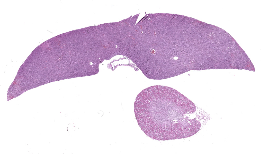
The liver was moderately enlarged, with rounded edges, beige to red in color and with an irregular bosselated surface.
The spleen was moderately enlarged, with multifocal to coalescing beige nodules.
Laboratory Results:
Not applicable.
Microscopic Description:
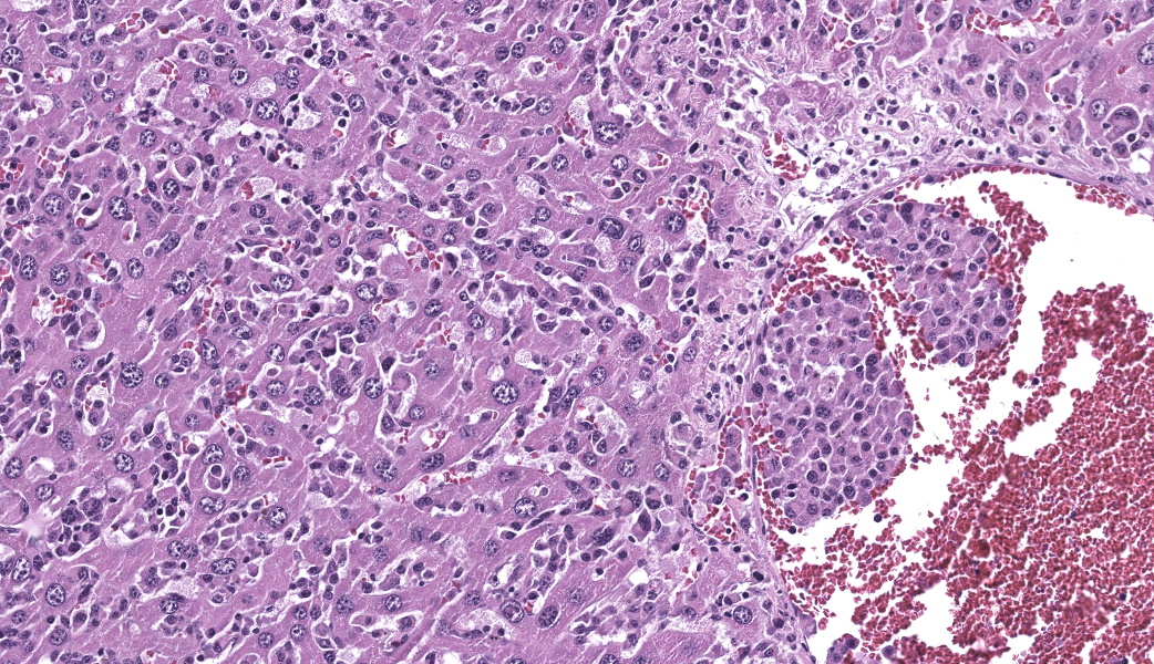
Liver: The normal hepatic architecture is markedly distorted and obscured by a multifocal to coalescing, highly infiltrative, poorly demarcated, unencapsulated, and densely cellular neoplasm. Neoplastic cells are arranged in sheets and small aggregates, infiltrating and expanding hepatic sinusoids and frequently displacing or replacing hepatocyte cords and stroma, thereby effacing normal lobular organization. The neoplastic cells are pleomorphic, with variably distinct cell borders, a moderate nuclear-to-cytoplasmic ratio, and a moderate amount of finely granular eosinophilic cytoplasm. Nuclei are round to ovoid with clumped chromatin and occasional prominent nucleoli. Multinucleated neoplastic cells are occasionally observed. There is marked anisocytosis and anisokaryosis. Mitotic figures are rare (1 in 10 high-power fields; 2.37 mm2). Neoplastic cells multifocally infiltrate the walls of larger vessels and frequently extend into the vascular lumina, forming intravascular aggregates (neoplastic emboli). Adjacent hepatocytes are frequently shrunken (atrophic), and surrounding sinusoids are either expanded by neoplastic cells or engorged with erythrocytes. Multifocal areas characterized by replacement of tissue with amorphous eosinophilic material and karyorrhectic debris (lytic necrosis) are also present, often associated with extravasated erythrocytes (hemorrhages) and occasional hemosiderin-laden macrophages. Scattered within hepatic sinusoids are small aggregates of small round cells with hyperchromatic nuclei, granulocytes and occasional megakaryocytes (extramedullary hematopoiesis).
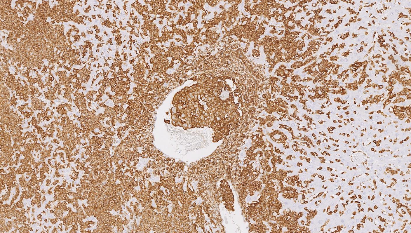
Immunohistochemistry reveals strong and diffuse cytoplasmic expression of Iba1 in the neoplastic cells. Lysozyme expression is evident in occasional neoplastic cells.
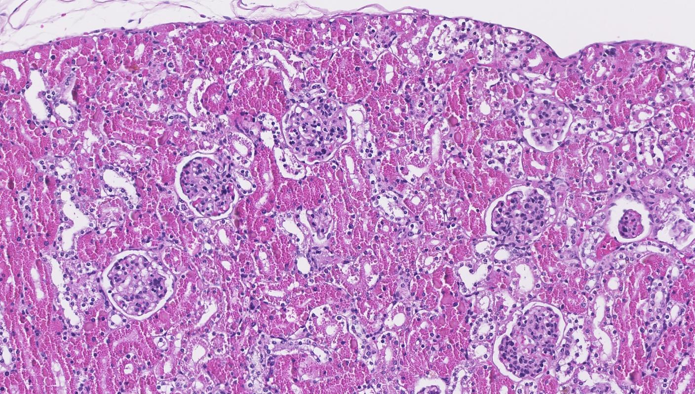
Kidney: The tubular epithelial cells of the proximal tubules are diffusely distended by numerous intensely eosinophilic, variably sized cytoplasmic granules (hyaline droplets). Occasional tubular epithelial cells contain fine intracytoplasmic brown pigment (presumed lipofuscin or hemosiderin). Rarely, tubular lumina contain homogeneous eosinophilic material (protein casts).
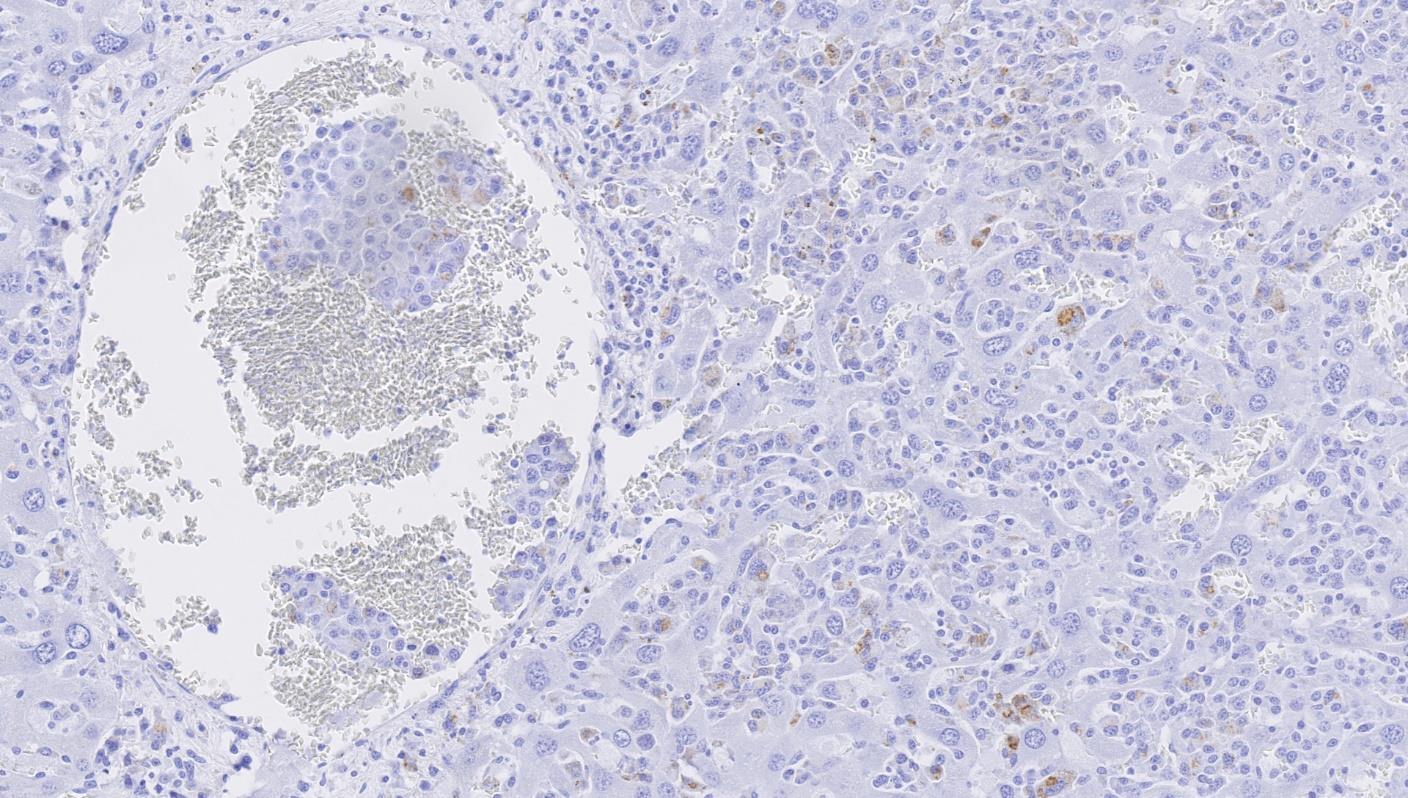
Immunohistochemistry reveals that the granules are diffusely positive for lysozyme. Occasional intravascular Iba1 positive cells are also evident.
Additional Findings (not present on the submitted slide): The neoplasm also extensively involved the spleen. Immunohistochemical staining for Iba1 also demonstrated intravascular aggregates of pleomorphic positive cells (neoplastic emboli) within vessels of the kidneys and lungs. Additionally, a fibrin thrombus was observed within the right ventricular chamber of the heart, adhering to the free wall and the septum.
Contributor's Morphologic Diagnoses:
Liver: histiocytic sarcoma with vascular wall invasion and neoplastic emboli.
Kidney: hyaline droplet accumulation in the tubular epithelial cells and rare protein casts.
Contributor's Comment:
Histiocytic sarcoma in mice is a relatively frequent and well-characterized hematopoietic neoplasm arising from the mononuclear phagocyte system, typically involving organs rich in resident macrophages, including the liver and spleen.2,6 In this case, the presence of multifocal to coalescing infiltrates of pleomorphic histiocytic cells in the liver, their intravascular localization, and strong immunolabeling with the histiocytic marker Iba1 are consistent with a diagnosis of histiocytic sarcoma.6
An associated and noteworthy finding in this case is the presence of numerous eosinophilic hyaline droplets in the proximal tubular epithelial cells of the kidneys. These droplets are considered a secondary, paraneoplastic change related to histiocytic sarcoma, due to the systemic overproduction of lysozyme - a major secretory product of neoplastic histiocytes.4,8
Hard and Snowden demonstrated that lysozyme-rich hyaline droplets are present in approximately 96% of rats and 55% of mice with histiocytic sarcoma, often correlating with tumor burden and distribution.4 Immunohistochemistry confirmed that these droplets were strongly positive for lysozyme (muramidase) and negative for alpha-1-antitrypsin, immunoglobulin, albumin, and alpha 2u-globulin.4 Luz and Murray further supported these findings across multiple mouse strains and emphasized that the visualization of hyaline droplets can be dependent on the eosin formulation used during staining, with alcoholic eosin providing superior contrast8.
From a mechanistic standpoint, overproduction of lysozyme by the neoplastic macrophages overwhelms the normal tubular degradation capacity. Lysozyme is a small (≅14 kDa) basic protein which is freely filtered and normally reabsorbed and catabolized in lysosomes of the tubular epithelial cells of the proximal renal tubule.4,9,10 Microscopically these appear as abundant, eosinophilic, homogenous cytoplasmic droplets (best seen in the P2 segment of proximal tubules).4 This parallels human monocytic leukemias where neoplastic cells produce lysozyme that similarly induce accumulation of protein droplets in the proximal tubule.9
The detection of these renal hyaline droplets should prompt careful evaluation for underlying histiocytic neoplasia in rodents, even in the absence of clinical suspicion. From a toxicologic pathology perspective, it is important to distinguish tumor-induced hyaline droplet accumulation from chemically-induced nephropathy, particularly in regulatory studies, to avoid misinterpretation of compound-related effects.2,4 Hyaline droplet accumulation in the tubular epithelial cells represents an important differential feature to distinguish this lesion from alpha 2u-globulin nephropathy, which occurs only in male rats and is chemically induced.2,4 Certain chemicals (e.g. hydrocarbons) bind alpha 2u-globulin and prevent its lysosomal degradation, so the protein accumulates in tubular lysosomes as spherical or crystalloid "hyaline droplets".3,4,11 These droplets are strongly indicative of male rat alpha 2u-globulin nephropathy: they are strongly positive for alpha 2u-globulin and absent in normal female rats or in mice (mice lack alpha 2u-globulin).4,7
Other neoplastic or degenerative causes of hyaline droplet accumulation in renal tubular epithelial cells are far less common but have been reported. Certain lymphomas or leukemias in mice have been associated with renal hyaline droplets.1 For example, Decker et al. described hyaline droplets in CD-1 mice with lymphoma.1 Mononuclear cell (large granular lymphocyte) leukemias in Fischer rats can have scattered pigmented granules in tubules (iron or lipofuscin, not lysozyme).4,5 In both the mouse and rat, any chronic severe proteinuric condition (e.g. glomerular disease or advanced chronic progressive nephropathy) can produce hyaline inclusions, but again the protein composition differs.2
Contributing Institution:
Laboratory for Animal Model Pathology
Institute of Veterinary Pathology
University of Zurich
Zurich, Switzerland
https://www.vetpathology.uzh.ch/de/Andere-Services/lamp.html
JPC Diagnosis:
- Liver: Histiocytic sarcoma.
- Kidney, proximal convoluted tubules: Hyaline droplet accumulation.
JPC Comment:
The contributor has provided an excellent review of the significance of hyaline droplet disease and its positive correlation with the presence of histiocytic sarcoma in affected individuals.
For a somewhat wider ranging review of this tumor (and specifically in C57Bl6 mice, the strain of the submitted individual in this case), the reader is referred to an excellent publication by Janke et al (2026).6 While a rare tumor in humans, this tumor is much more common in mice, including the C57Bl6 strain, where in various studies it ranges from 2-34.7%.6 It is a tumor of many different morphologies, which has resulted in its publication as a wide range of tumors, including lymphoma, Schwannoma, endometrial sarcoma (spindle cells is a morphologic hallmark of this tumor) and often many others.6
Histiocytic sarcoma in mice is renowned for morphologic heterogeneity; however, round cells and spindle cells are typically present in the same tumor. In the review by Janke, 39% of tumors demonstrated a range of cellular pleomorphism, and multinucleated cells were seen in 21% of cases. Intravascular involvement, as seen in this case, was present in 61% of tumors in that study and areas of necrosis was present in 42%.
Additionally, to further complicate the diagnostic picture (at least on HE review), this tumor often coexists (and may be commingled) with murine lymphoma or leukemia (12% of the animals in the Janke study).6 B-cell tumors predominated at a rate of 88%.6
Extramedullary hematopoiesis is a common finding in this tumor, both within the tumor and in other organs which may lack tumor cells (typically in affected animals, the spleen is rife with EMH, but rarely the tumor itself). In the Janke study, EMH was seen in 75% of tumors.6 Splenic hematopoiesis was first reported by Frith et al in 199012, and hepatic hematopoiesis was reported three years later by Lacroix-Triki et al.12 While the definitive connection between histiocytic sarcoma and extramedullary hematopoeisis (both within the tumor itself and in remote locations) remains elusive, macrophages are well known for their vital roles in erythropoiesis, which include support of hematopoetic stem cells as well as the removal of senescent stem cells, and creating and regulating a specialized microenvironment for both erythroid and megakaryocyte precursors.14
References:
- Decker JH, Dochterman LW, Niquette AL, Brej M. Association of Renal Tubular Hyaline Droplets with Lymphoma in CD-1 Mice. Toxicol Pathol. 2012;40(4):651-655.
- Frazier KS, Seely JC, Hard GC, et al. Proliferative and Nonproliferative Lesions of the Rat and Mouse Urinary System. Toxicol Pathol. 2012;40(4_suppl):14S-86S.
- Hamamura M, Oshikata T, Katoku K, Tsuchitani M, Yamaguchi R. Two types of deposits, hyaline droplets and eosinophilic bodies, associated with alpha 2u-globulin accumulation in the rat kidney. J Toxicol Pathol. 2017;30(4):275-282.
- Hard GC, Snowden RT. Hyaline Droplet Accumulation in Rodent Kidney Proximal Tubules: An Association with Histiocytic Sarcoma. Toxicol Pathol. 1991;19(2):88-97.
- Ishmael J, Dugard PH. A review of perchloroethylene and rat mononuclear cell leukemia. 2006;45(2):178-184.
- Janke LJ, Rehg JE. The many faces of mouse histiocytic sarcoma in C57BL/6J mice. Vet Pathol. 2023;60(4):443-460.
- Lehman-McKeeman LD, Caudill D. Biochemical basis for mouse resistance to hyaline droplet nephropathy: Lack of relevance of the alpha 2u-globulin protein superfamily in this male rat-specific syndrome. Toxicol Appl Pharmacol. 1992;112(2):214-221.
- Luz A, Murray AB. Hyaline Droplet Accumulation in Kidney Proximal Tubules of Mice with Histiocytic Sarcoma. Toxicol Pathol. 1991;19(4-2):670-671.
- Santoriello D, Andal LM, Cox R, D'Agati VD, Markowitz GS. Lysozyme-Induced Nephropathy. Kidney Int Rep. 2017;2(1):84-88.
- Singh G, Katyal SL, Brown WE, Collins DL, Mason RJ. Pulmonary Lysozyme-A Secretory Protein of Type II Pneumocytes in the Rat. 1988;138(5):1261-1267.
- Urinary System Kidney, Renal Tubule - Accumulation, Hyaline Droplet, https://ntp.niehs.nih.gov/atlas/nnl/urinary-system/kidney/RenalTubule-AccumulationHyalineDroplet. Nonneoplastic Lesion Atlas - National Toxicology Program.
- Frith CH (1990). Histiocytic sarcoma, mouse. In: Monographs on Pathology of Laboratory Animals. Hemopoietic system, Jones TC,Ward JM, Mohr U, Hunt RD (eds). Springer-Verlag, Berlin, Heidelberg, pp 58-65.
- Lacroix-Triki M, Lacoste-Collin L, Jozan S, Charlet JP, Caratero C, Courtade M. Histiocytic sarcoma in C57BL/6J female mice is associated with liver hematopoiesis: review of 41 cases. Toxicol Pathol. 2003 31(3):304-9.
- Huang H, Gao M, Vinchi F, An X, Li W, Wang Y. Macrophages in Hematopoiesis and Related Blood Diseases. Genomics Proteomics Bioinformatics. 2025 23(6):qzaf112. doi: 10.1093/gpbjnl/qzaf112.