Signalment:
Gross Description:
Histopathologic Description:
Morphologic Diagnosis:
Condition:
Contributor Comment:
To date, only female Halicephalobus have been found in tissue samples.(10) While the route of infection and pathogenesis of disease are poorly understood, it is suspected that the organism gains entry via existing wounds. In some cases, viable Halicephalobus organisms have been detected in sperm and urine;(9) while no transmission via this route has been proven, it is another possible source of infection. Organisms access the central nervous system via blood vessels, and cause necrosis and inflammation due to migration through the tissue.
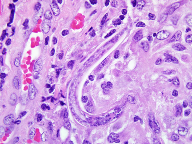
Under light microscopy, adult females are typically approximately 20 μm in diameter and 350 μm long, with a thin, smooth cuticle and tapered, pointed tail. They have platymyarian-meromyarian musculature, a rhabditiform esophagus, and an intestine lined by low cuboidal cells. Larvae are approximately 10 μm in diameter, with tapered, pointed tails. Typically, tissue sections have numerous parasites; this may be explained by the fact that females are parthenogenetic and thus can produce offspring in the absence of males.
Other rule outs for rhabditoid parasites in tissue sections include Cephalobus spp., Strongyloides westeri, and Pelodera strongyloides.(12) Cephalobus spp. can be differentiated from Halicephalobus spp. by examination of the tail, which is blunt in Cephalobus.(6) Strongyloides westeri have alae, which Halicephalobus spp. lack.(6) Pelodera strongyloides also have two lateral alae, as well as two lateral cords noted by two densi nuclei.(7)
JPC Diagnosis:
Conference Comment:
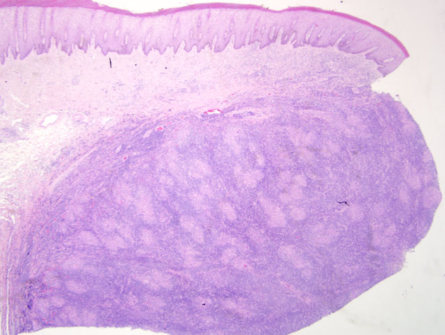
In addition to the histomorphologic differences among the various nematodes, clinicopathologic presentation may assist with differentiating them. Although H. gingivalis dermatitis may occur anywhere on the skin of horses, it generally localizes near or on the prepuce; lesions tend to be papules or nodules that are well circumscribed, measure 0.5 cm to 8 cm in diameter, and are often ulcerated.(14) In contrast, Pelodera dermatitis is characterized by papules, pustules, ulcers, crusts, alopecia, and scales on areas of the skin that typically have contact with damp soil and decaying organic matter, such as the limbs, ventral thorax, and abdomen;(14) pruritis is usually moderate to intense.
We thank Dr. Christopher Gardiner, Consulting Parasitologist for the AFIPs Department of Veterinary Pathology, for his study and diagnostic commentary for this case.
References:
2. Bryant U, Lyons E, Bain F, Hong C. Halicephalobus gingivalis-associated meningoencephalitis in a Thoroughbred foal. J Vet Diagn Invest. 2006;18:612-615.
3. Br+â-¦jer J, Parsons D, Linder K, Peregrine A, Dobson H. Halicephalobus gingivalis encephalomyelitis in a horse. Can Vet J. 2000;41:559-561.
4. Dunn D, Gardiner C, Dralle K, Thilsted J. Nodular granulomatous posthitis caused by Halicephalobus (syn. Micronema) sp. in a horse. Vet Pathol. 1993;30:207-208.
5. Ferguson R, van Dreumel T, Keystone J, et al. Unsuccessful treatment of a horse with mandibular granulomatous osteomyelitis due to Halicephalobus gingivalis. Can Vet J. 2008;49:1099-1103.
6. Greiner E, Mays M, Smart GJ, Weisbrode S. Verminous mastitis in a mare caused by a free-living nematode. J Parasitol. 1991;77:320-322.
7. Gutierrez Y. Diagnostic Pathology of Parasitic Infections with Clinical Correlations. 2nd ed. New York, NY: Oxford University Press; 2000:307.
8. Isaza R, Schiller C, Stover J, Smith P, Greiner E. Halicephalobus gingivalis (Nematoda) infection in a Grevy's zebra (Equus grevyi). J Zoo Wildl Med. 2000;31:77-81.
9. Kinde H, Mathews M, Ash L, St Leger J. Halicephalobus gingivalis (H. deletrix) infection in two horses in southern California. J Vet Diagn Invest. 2000;12:162-165.
10.Muller S, Grzybowski M, Sager H, Bornand V, Brehm W. A nodular granulomatous posthitis caused by Halicephalobus spp. in a horse. Vet Dermatol. 2008;19:44-48.
11.Rames D, Miller D, Barthel R, et al. Ocular Halicephalobus (syn. Micronema) deletrix in a horse. Vet Pathol. 1995;32:540-542.
12.Rashmir-Raven A, Black S, Rickard L, Akin M. Papillomatous pastern dermatitis with spirochetes and Pelodera strongyloides in a Tennessee Walking Horse. J Vet Diagn Invest. 2000;12:287-291.
13.Ruggles A, Beech J, Gillette D, Midla L, Reef V, Freeman D. Disseminated Halicephalobus deletrix infection in a horse. J Am Vet Med Assoc. 1993;203:550-552.
14.Scott DW, Miller WH, Jr. Equine Dermatology. St. Louis, MO: Elsevier Ltd; 2003:366-369.
15.Shibahara T, Takai H, Shimizu C, Ishikawa Y, Kadota K. Equine renal granuloma caused by Halicephalobus species. Vet Rec. 2002;151:672-674.