Signalment:
8-day-old, male, miniature horse (foal),
Equus ferus caballusPresented for complete necropsy following natural death. It had a reported clinical history of impaction.
Gross Description:
The foal had a body condition score of 3/5 and was in fair post-mortem condition. The adipose tissue throughout the carcass was pale yellow (icterus). The liver contained numerous pinpoint pale tan and red-black foci throughout. Upon opening the elbow joints, minimal amounts of yellow, strand-like material was adherent to the cartilaginous surface of the humeral condyles and the radial fossa (fibrinosuppurative arthritis). The stomach contained a moderate amount of pale yellow, pasty material (digested milk), while the small intestines and cecum contained mild to moderate amounts of dark gray, opaque, viscous digesta. The large and small colon contained moderate amounts of yellow pasty feces throughout.
Histopathologic Description:
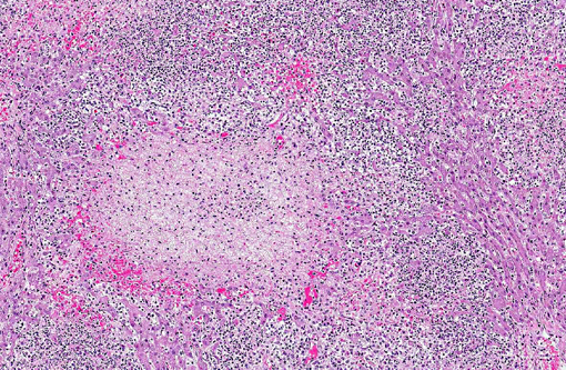
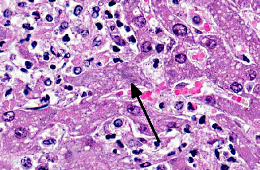
Liver: The hepatic parenchyma is replaced by multifocal to coalescing round to oval aggregates of hepatic necrosis and inflammation. These areas are characterized by a central area of hepatocytes undergoing coagulative and liquifactive necrosis admixed with homogenous eosinophilic fibrillar material (fibrin) and neutrophils undergoing necrosis. The areas of hepatocellular necrosis are often surrounded by a dense layer of karyorrhectic and karyolytic nuclear debris from necrotic neutrophils. Hepatocytes can be observed at the periphery of these lesions that contain intracytoplasmic aggregates of rod-shaped bacteria.
Morphologic Diagnosis:
Marked acute multifocal random necrosuppurative hepatitis with intracytoplasmic bacilli
Condition:
Tyzzer's disease
Contributor Comment:
Tyzzers disease was first described in laboratory rodents, but has been reported in a number of species as a naturally occurring disease.(3) The causative organism is
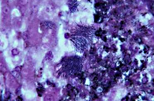
Clostridium piliforme, formerly known as
Bacillus piliformis. The organism is a gram-negative, obligate intracellular, spore-forming rod. Exposure is likely through the fecal-oral route with systemic dissemination taking place after infection establishes in the intestines. The liver and myocardium are primarily affected. Necrotizing enteritis can be seen in some laboratory animal species, most notably rodents and rabbits, with the distal small intestine primarily affected. Silver stains (e.g. Warthin-Starry or Gomoris methanamine silver) are useful in highlighting the bacteria.(3) Immunohistochemical or immunofluorescence staining can also be used to confirm the diagnosis.(1)
Foals from 7 to 42 days of age are primarily affected(1) and immunocompromised animals are predisposed to infection. The organism is not considered highly contagious and animals are typically affected sporadically in a given population. Infected animals can be found dead or may present for acute onset of non-specific clinical signs such as pyrexia, lethargy, anorexia and icterus. Typically elevations of hepatocellular leakage enzymes and hyperbilirubinemia take place.(3) Treatment of animals with confirmed disease is typically unrewarding.
JPC Diagnosis:
Liver: Hepatitis, necrotizing and suppurative, multifocal to coalescing, severe, with intracytoplasmic filamentous bacilli.
Conference Comment:
The contributor provides a good summary of
C. piliforme, an atypical member of the genus
Clostridium. Other members of the clostridia are large, Gram positive spore forming bacteria with straight or slightly curved morphology, in contrast the filamentous, Gram negative spore forming
C. piliforme.(2) Further differentiating
C. piliforme from other clostridia is the fact that it does not possess characteristics that allow its inclusion into one of the three general categories of the other pathogenic members of the genus. These categories of clostridia are neurotoxic (
C. tetani, C. botulinum types A-G), histotoxic (
C. chauvoei, C. septicum, C. novyi types A and B,
C. perfringens type A,
C. sordellii, C. hemolyticum), and enteropathogenic/enterotoxemia-producing (
C. perfringens types A-E,
C. difficile, C. colinum, C. spiroforme).(2)
Conference participants discussed the importance of recognizing both the pattern of necrosis (i.e. multifocal, random) as well as the inflammatory component in this section, and discussed the differential diagnosis for such lesions. The conference moderator provided the following table of common causes of bacterial hepatitis in various domestic animal species:
| Organism | Species Affected |
| Listeria monocytogenes | Neonates: ruminants, foals, piglets |
| Campylobacter fetus | Lambs, foals, piglets |
| Actinobacillus equilli/suis | Foals/piglets |
| Yersinia pseudotuberculosis | Lambs, dogs, cats |
| Francisella tularensis | Lambs, cats |
| Mannheimia hemolytica/Histophilius somni | Lambs |
| Salmonella spp. | Various |
| Clostridium piliforme | Foals, dogs |
| Nocardia/Mycobacterium | Various |
References:
1. JJ Van Der Lugt. Tyzzers disease. In : JAW Coetzer, TC Tustin, ed.
Infectious diseases of livestock, vol. 3. Oxford University Press, 2004. pp. 2089-2091.
2. Quinn PJ, Markey BK, Leonard FC, FitzPatrick ES, Fanning S, Hartigan PJ. Clostridium species. In:
Veterianry Microbioogy and Microbial Disease. 2nd edition. Ames, IA: Wiley-Blackwell; 2011: Kindle edition.
3. MJ Stalker, MA Hayes. Liver and biliary system In: Maxie MG , ed.
Jubb, Kennedy and Palmers Pathology of Domestic Animals. 5th ed. Vol 2. Philadelphia, PA: Elsevier; 2007:356.