Joint Pathology Center
Veterinary Pathology Services
Wednesday Slide Conference
2017-2018
Conference 25
May 9th, 2018
CASE I: 17B-0101 (JPC 4102436).
Signalment: 9-year-old, neutered male, mixed breed (Canis familiaris), canine.
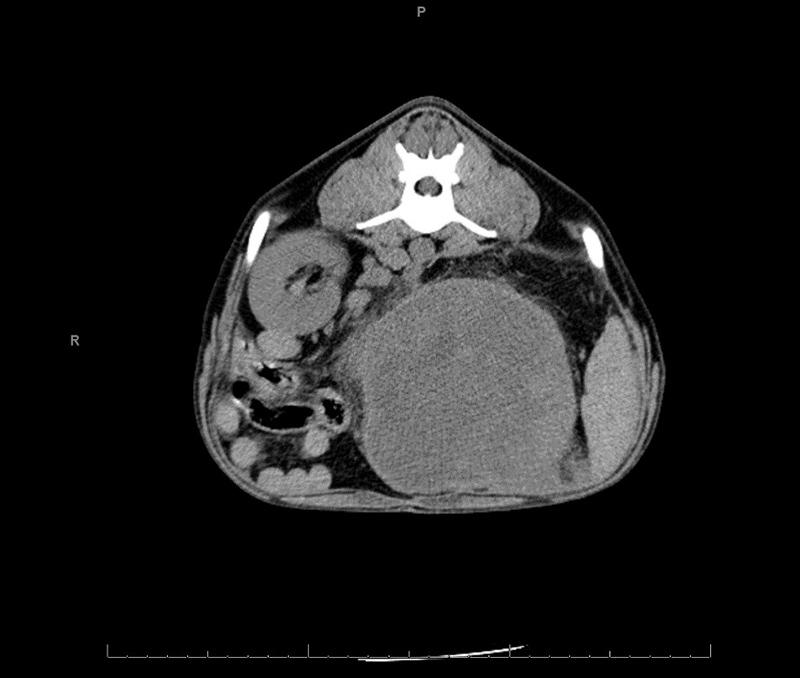
History: The dog had an approximately 3 week history of hematuria which improved but did not resolve with antibiotics. Abdominal ultrasound and CT scan revealed a mass lesion involving the left kidney. Cytologic evaluation of the mass lesion revealed spindle cells of uncertain origin (reactive vs. neoplastic). The left adrenal gland was not visualized via imaging or during surgery. Surgical findings reported white streaking along the mesentery suggestive of lymphatic invasion.
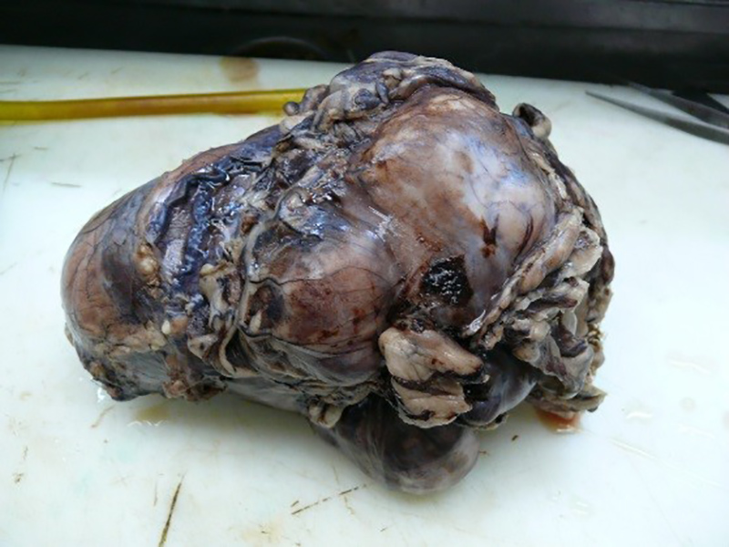
Gross Pathology: Received is a 20 x 11 x 10 cm multilobulated mass lesion with multifocally adhered omentum. The kidney itself is not apparent until sectioning. On cut section, the mass lesion is soft, multilobular, red and tan with a thin rim of remnant kidney representing approximately 10% of the submitted tissue. The adrenal gland is not apparent.
Laboratory results: None provided.
Microscopic Description: Left kidney: Where anatomic borders are apparent, the leading edge of the mass lesion is necrotic and thus, poorly demarcated. Regions of the border are composed of fibrous connective tissue which could either represent tumor capsule or could represent reactive fibrosis of the kidney.
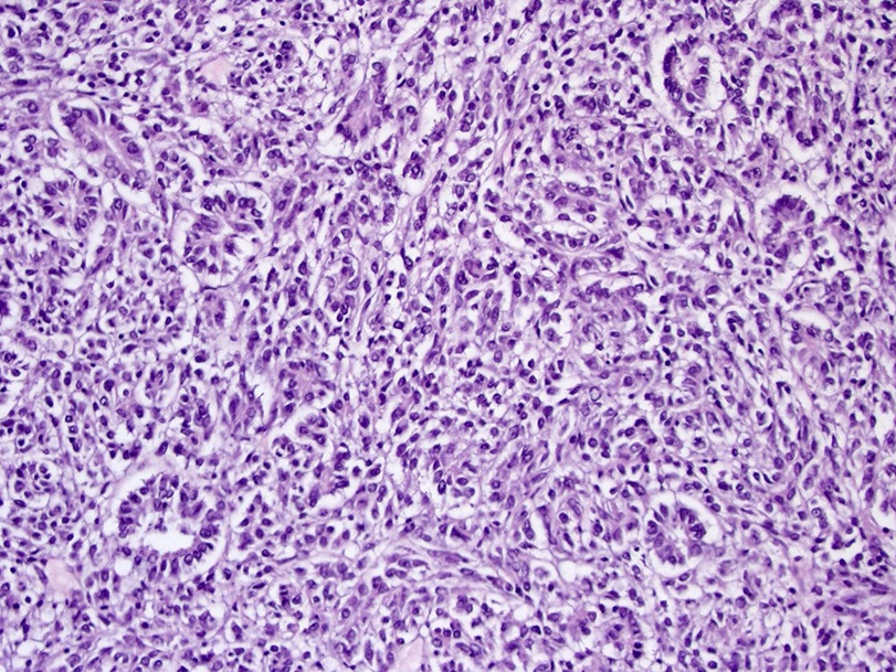
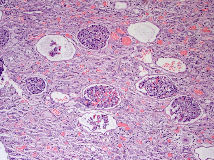
The mass lesion is composed of multiple lobules of spindle cells usually arranged in streams, but occasionally forming tubules, acini, and rosettes all of which have similar cellular morphology. The fibrovascular stroma is fine. Neoplastic cells have indistinct cell borders, moderate amounts of eosinophilic cytoplasm, and round to oval nuclei with coarsely stippled chromatin. Mitotic figures average 8 in 10 high powered fields examined. Anisocytosis and anisokaryosis are moderate. Multifocally there is abundant tumor necrosis, variably coagulative and lytic, with multifocal hemorrhage. The tumor is separated from the surgical margin by a single layer of spindle cells. There is no evidence of vascular invasion. The adrenal gland is not histologically apparent.
Contributor’s Morphologic Diagnosis: Left kidney: Nephroblastoma
Contributor’s Comment: Nephroblastoma is the most common primary renal tumor of swine, chickens and fish, the fifth most common primary renal tumor in dogs, and the fourth most common primary renal tumor in cats.7 A tumor with similar morphology also occurs in the thoracolumbar spinal column of young dogs.7
The age distribution in veterinary species follows that in people where it is an embryonal, childhood tumor. In this case and other reported cases in dogs, the tumor is discovered in adulthood but is suspected to either be present from a young age or to arise from remnant nephrogenic rests later in life.2,7
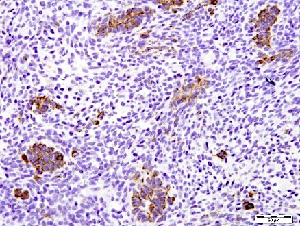
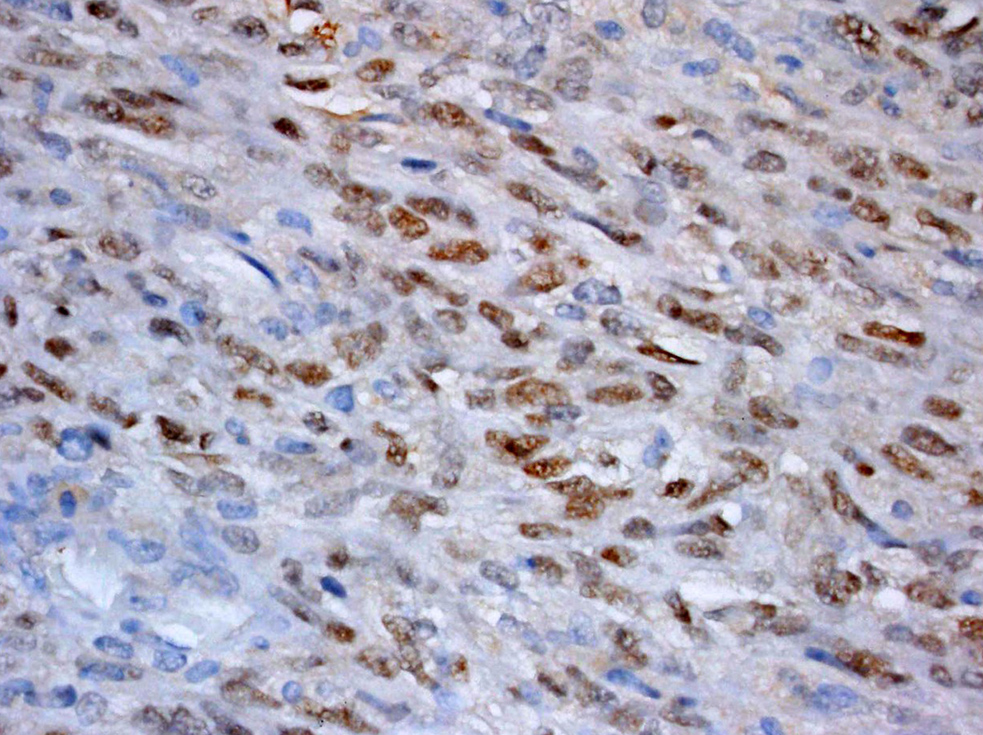
Despite Dr. Meuten’s commentary that “searching sufficient sections will eventually identify a few [glomeruloid structures] that are worthy of the diagnosis or a photograph,” numerous DVM and MD pathologists at our institution were unable to do so. Because this tumor did not have classic features of primitive glomeruli, the slides were reviewed with local collaborating MD pathologists with expertise in embryonic renal tumors. Positive immunohistochemical staining for Wilms’ tumor 1 (WT1) confirmed the diagnosis of nephroblastoma and ruled out their differential diagnosis of synovial cell sarcoma which, in humans, can be a primary mesenchymal renal tumor diagnosed via morphology together with specific chromosomal translocations.7
Metastatic risk is considered high in dogs4,7, but primary source data for this assertion is difficult to confirm, and sample sizes are usually small in published studies. In a 2006 retrospective paper1 detailing clinical outcome of dogs with primary renal neoplasia, only 5 of 82 dogs had the diagnosis of nephroblastoma. Of those, one reported metastatic disease at the time of diagnosis and three of four from which follow-up was available had metastatic disease at the time of death. The spindle cell variant is also reported to be of higher metastatic potential4 also without a reference to primary source data. The tumor in the submitted case progressed quickly both locally and distantly in spleen and abdominal lymph nodes within 6 weeks of resection. The blastemal predominant morphology of this tumor has been reported in at least two other single case reports in dogs9,10, the former representing a benign variant.
Cianciolo and Mohr report tumors with more differentiated tubular and glomerular structures have a better prognosis than those with anaplasia and sarcomatous features2 although the primary source for this statement is unclear. Diagnosis in this case was challenging in that the characteristic feature of glomeruloid structures were not apparent.
JPC Diagnosis: Kidney: Nephroblastoma, mixed breed (Canis familiaris), canine.
Conference Comment: Nephroblastomas are a generally disorganized mixture of three components: epithelial, mesenchymal, and blastemal which arise from the metanephrogenic renal blastema. In different tumors and even different areas within the same tumor one or more of those components can predominate. In humans, the causative factor is mutation of the tumor suppressor gene Wilm’s tumor-1 (WT-1) which is located on chromosome 11p13. A genetic cause in animals has not yet been identified. Grossly, nephroblastomas are most often unilateral and isolated to one pole of the kidney, encapsulated, multilobulated, white to tan, and firm with spongy/cystic areas and foci of necrosis and hemorrhage. Microscopically, the epithelial component often forms abortive tubules or primitive glomeruli within tubules surrounded by the mesenchymal components which is organized in long interlacing streams and can differentiate into muscle, fibrous, adipose, cartilage or bone tissues. The blastemal component is often found in clumps between the epithelial and mesenchymal components and are small cells with high nuclear to cytoplasmic ratios and round, hyperchromatic nuclei.2,7
As mentioned above, it has been described that tumors with mesenchymal differentiation often exhibit frequent metastasis.4 There has been recent publication of a blastemal-predominant canine renal nephroblastoma that had gingival metastasis which suggests that all types of nephroblastomas have metastatic potential. In dogs, a common presentation occurs not in the kidney, but in the thoracolumbar spinal cord, arising from remnants of metapnephric rests located between the dura mater and spinal cord. This condition is most commonly observed in young German Shepherd Dogs and is also called ectopic nephroblastoma. Additionally, there have been rare reports of multifocal spinal cord involvement which may represent metastasis or multifocal generation of the tumor.3,11 Traditional renal nephroblastomas have been identified in all species but occur with greatest frequency in chickens and pigs.2 Where in chickens, they are often associated with avian leukosis virus.8 In fish (Japanese eel and Rainbow trout) and rats (Sprague-Dawley subline) nephroblastomas are induced via carcinogens.5,6
Contributing Institution:
University of Wisconsin Madison
School of Veterinary Medicine
Pathobiological Sciences
References:
- Bryan JN, Henry CJ, Turnquist SE, Tyler JW, et al. Primary renal neoplasia of dogs. 2006; 20(5):1155–1160.
- Cianciolo R, Mohr C. Urinary system. In: Maxie MG, ed. Jubb, Kennedy & Palmer's Pathology of Domestic Animals. Vol 2. 6th ed. St. Louis, MO: Elsevier; 2016:376-464.
- Henker LC, Bianchi RM, Vargas TP, DeOliveira EG, et al. Multifocal spinal cord nephroblastoma in a dog. J Comp Pathol. 2018;158:12-16.
- Ladanyi M. Fusions of the SYT and SSX genes in synovial sarcoma. Oncogene. 2001; 20(40):5755-5762.
- Lombardini ED, Hard GC, Harshbarger JC. Neoplasms of the Urinary Tract in Fish. Vet Pathol. 2014;51:1000-1012.
- Mesfin GM. Intralobar nephroblastematosis: precursor lesions of nephroblastoma in the Sprague-Dawley rat. Vet Pathol. 1999;36:379-90.
- Meuten D, Meuten T. Tumors of the urinary system. In: Meuten DJ, ed. Tumors in Domestic Animals. 5th ed. Ames, IA: John Wiley & Sons; 2017:632-688.
- Nair V, Fadly AM. Neoplastic diseases: Leukosis/sarcoma group. In: Swayne DE, ed. Diseases of Poultry. 13th ed. Ames, IA: Wiley-Blackwell Publishing; 2013:578-582.
- Simpson RM, Gliatto M, Casey HW, Henk WG. The histologic, ultrastructural, and immunohistochemical features of a blastema-predominant canine nephroblastoma. Vet Pathol. 1992; 29(3):250-253.
- Soldati S, Radaelli E, Mazzuti A, Scanziani E. Congenital mesoblastic nephroma in a young basset hound dog. J Small Anim Pract. 2012; 53:709–713.
- Terrell SP, Platt SR, Chrisman CL, et al. Possible intraspinal metastasis of a canine spinal cord nephroblastoma. Vet Pathol. 2000;37:94-97.