Signalment:
10-year-old, spayed female mixed breed, canine (
Canis familiars).This animal had a cutaneous mass removed from the inter-scapular region three years previously which was diagnosed as a malignant pilomatricoma. The animal presented recently with labored breathing and was euthanized due to numerous pulmonary soft-tissue opacities radiographically. Portions of lung were submitted for histopathology.
Gross Description:
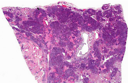
In multifocal to coalescing regions, there is extensive replacement of the pulmonary parenchyma by soft, pale tan to red, variably necrotic masses.Â
Histopathologic Description:
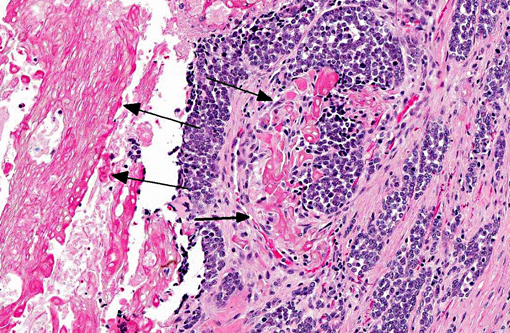
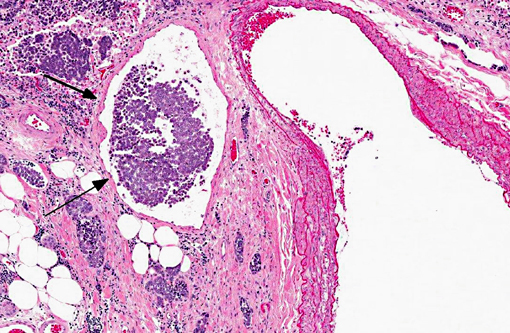
The normal pulmonary architecture is extensively effaced by neoplastic epithelial cells, necrosis, and debris. The basaloid cells are arranged in coalescing cords and clusters supported by a fibrovascular to inflammatory stroma. The cells have indistinct cell borders and small amounts of eosinophilic cytoplasm and central round to ovoid nuclei with coarse, irregular chromatin and prominent nucleoli. There are scattered foci of squamous differentiation. The cells exhibit moderate to marked anisocytosis and anisokaryosis. Mitoses are common and range from 3-10 per 40X field. The cell clusters occasionally have central necrosis or central keratinized "ghost cells." Tumor emboli are common in vessels and lymphatics.
Morphologic Diagnosis:
Lung: Metastatic pilomatricoma.
Condition:
Malignant pilomatricoma
Contributor Comment:
Pilomatricoma is a benign cutaneous tumor that arises from the germinative cells of the follicular matrix, or hair bulb exhibiting only matrical differentiation.(1,2,4) It is an uncommon neoplasm of dogs and comprises approximately 1% of canine skin tumors.(1,3) Pilomatricomas most commonly occur on the back, neck, thorax, and tail. The tumors are generally well-delineated, firm, multilobular intradermal masses that often contain areas of grey-white chalky material on cut section.(2) Young, adult dogs are typically affected. Poodles, Kerry Blue Terriers, Old English Sheepdogs, Soft-coated Wheaten Terriers, and other breeds with continuously growing coats are over-represented which is believed to be related to the continuously-growing coat and subsequently greater numbers of mitotically active anagen follicles in these breeds.(1,4)
The histological features include a well-delineated dermal and/or subcutaneous epithelial tumor composed of multiple, variably sized cystic structures supported by a collagenous stroma. The structures are lined with several layers of basaloid keratinocytes which resemble the matrix cells of the anagen hair bulb with a moderate to high mitotic activity. Scattered squamous differentiation can be seen. As the neoplastic epithelial cells differentiate towards the center of the lesion they exhibit matrical keratinization (ghost or shadow cells) and the center of the cysts accumulate large aggregates of ghost cells and debris. The central debris can degenerate and become mineralized with foci of osseous metaplasia.(2,4)
Malignant pilomatricomas are rare and can be distinguished from the benign variety by their poorly delineated, infiltrative nature, increased mitotic activity, and increased ratio of basaloid cells to keratinized ghost cells. Lymphatic invasion is often evident along the periphery of the lesion. Metastasis has been reported to local lymph nodes, bone, and lung.(1,2,3)
JPC Diagnosis:
Lung: Metastatic pilomatricoma.
Conference Comment:
Recently, Martano et al. reported on the histopathological and immunophenotypic characteristics of malignant pilomatricoma with metastasis to bone in an 11-year-old mongrel dog.(5) Malignant pilomatricoma was diagnosed in an ulcerated mass from the dogs neck and a nodular bone mass, based on the histopathological features of irregularly-shaped, lobulated islands of basaloid cells undergoing abrupt keratinization to shadow cells, a high mitotic rate, and nuclear atypia, as well as metastasis to bone. Anti β-catenin immunohistochemistry revealed strong nuclear immunoreactivity of neoplastic basaloid cells, with few transitional cells exhibiting membrane-bound immunoreactivity. In humans, although the extent of membrane, cytoplasmic and nuclear expression of β-catenin varies in most skin tumors, basaloid cells in pilomatricoma and malignant pilomatricoma usually demonstrate intense diffuse nuclear immunoreactivity with β-catenin antibody. Furthermore, pilomatricoma in humans are often associated with deregulation of the Wnt/β-catenin pathway due to mutations in the
CTNNB1 gene. In normal cells, β-catenin can be located at the cell surface where it links E-cadherin to the actin cytoskeleton to create cell-to-cell junctions. Cytosolic β-catenin is phosphorylated by the APC-AXIN-GSK3β destruction complex, and subsequently destroyed via ubiquitination (known as the APC/ β-catenin pathway).(6) Wnt signaling blocks the destruction complex, allowing β-catenin to accumulate and translocate to the nucleus, where it complexes with transcription factors that up-regulate transcription of c-MYC, cyclin D1, and other genes that increase cellular proliferation. Dysregulation of the APC/ β-catenin pathway has been found to play a role in the development of several neoplasms, including colon tumors and hepatocellular carcinomas in addition to pilomatricomas.(5,6) Based on the findings in this study, Martano et al. suggest that, similar to humans, β-catenin is involved in the pathogenesis of malignant pilomatricoma in dogs, and therefore may be a useful diagnostic marker.(5)
References:
1. Carroll EE, Fossey SL, Mangus LM, Carsillo ME, Rush LJ, McLeod CG, Johnson TO. Malignant pilomatricoma in 3 dogs.Â
Vet Path. 2010;47(5):937-943.
2. Goldschmidt MH, Hendrick MJ. Tumors of the skin and soft tissue: Pilomatricoma and malignant pilomatricoma. In:
Tumors in Domestic Animals. 4th ed. Ames, Iowa: Iowa State Press; 2002:61-63.
3. Gross TL, Ihrke PJ, Walder EJ, Affolter VK. Malignant pilomatricoma. In:
Skin Diseases of the Dog and Cat, Clinical and Histopathologic Diagnosis. 2nd ed. Ames, Iowa: Blackwell Publishing; 2005:637-638.
4. Gross TL, Ihrke PJ, Walder EJ, Affolter VK. Pilomatricoma. In:
Skin Diseases of the Dog and Cat, Clinical and Histopathologic Diagnosis. 2nd ed. Ames, Iowa: Blackwell Publishing; 2005:624-625.
5. Martano M, et al. Malignat pilomatricoma with multiple bone metastases in a dog: Histological and immunohistochemical study.Â
Exp Ther Med. 2013;5(4):10051008.
6. Stricker TP, Kumar V. Neoplasia. In: KumarV, Abbas AK, Fausto N, Aster JC, eds.Â
Robbins and Cotran Pathologic Basis of Disease. 8th ed. Philadelphia, PA: Saunders Elsevier;2010:292-294.Â