Signalment:
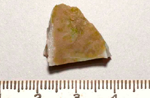
Gross Description:
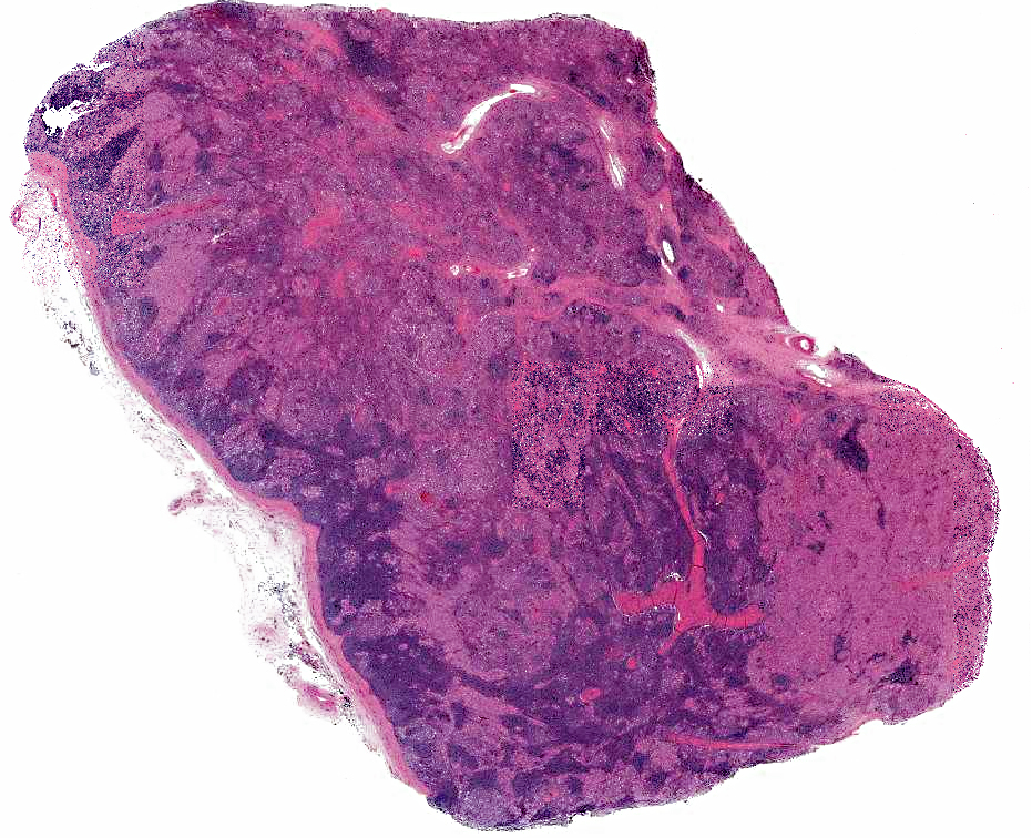
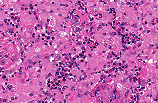
Histopathologic Description:
Morphologic Diagnosis:
Condition:
Contributor Comment:
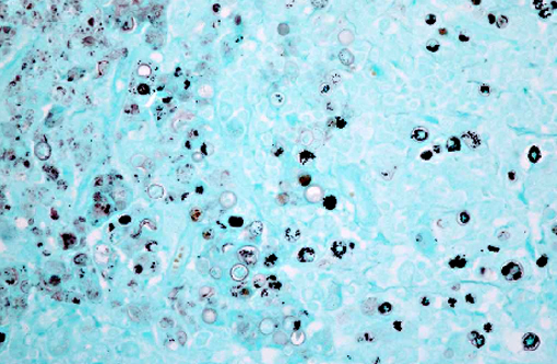
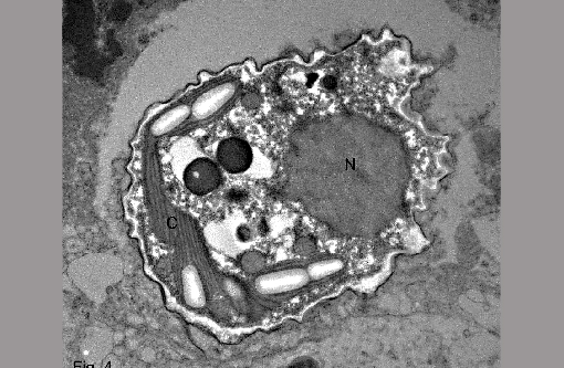
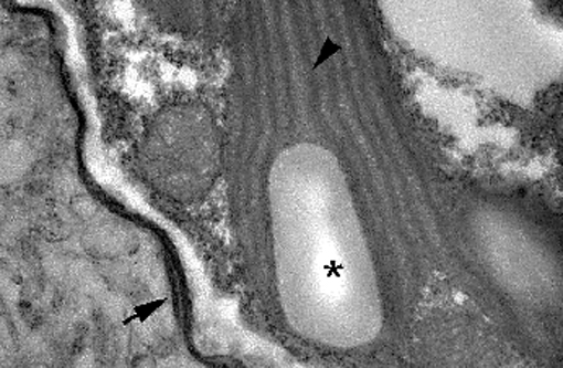
Green algal infections have been described most commonly in sheep and in cattle, with individual case reports in a dog, man and several species of wild mammals.(1,4,5) The algae are found commonly throughout the world and infections have been associated with stagnant water or pasture irrigated with raw sewage.(5) Disease varies from subclinical with a localized lymphadenitis to severe clinical disease and systemic lesions.(1,2,5) Underlying immunosuppression or an overwhelming infectious dose are possible predisposing factors to causing disease by this ubiquitous organism.(5) Our case is consistent with one of the largest reports (a group of 8 cattle cases) in which green algae induced lymphadenitis was identified through postmortem slaughter inspection.(6) Green algal infections are also identified several times a year in cattle by the USDA Food Safety Inspection Service pathology laboratory, which receives samples from federally inspected slaughter facilities (S Hafner, personal communication). Based on the lesion and organism morphology on HE stained slides, the primary differential diagnoses include various types of green algae and Prototheca spp. Protothecosis occurs more commonly than disease by green algae and Prototheca spp. are achlorous, which is a key criteria for differentiating green algae from Prototheca spp.(1,5) Chandler et. al.(1) uses three criteria for differentiating Chlorella (a type of green algae) from Prototheca spp. and for making a presumptive diagnosis. In chlorellosis there are: 1) grossly visible green discoloration of the tissues due to the presence of chlorophyll in the algae, 2) spherical algal cells, with an average diameter of 9 microns that exhibit endosporulation, and 3) algal cells that have abundant large cytoplasmic granules that are strongly positive with PAS, GF, and GMS stains.(1) Other methods used to differentiate the two include impression smears of fresh tissue and transmission electron microscopy, both of which are based on the presence of numerous chlorophyll/chloroplasts.(1,5) It is important to remember that the green color of the Chlorella algae is lost during tissue fixation and processing.(1)
JPC Diagnosis:
Conference Comment:
In addition to Chlorella and Prototheca, other pathogens that reproduce asexually by endosporulation include Rhinosporidium seeberi and Coccidioides immitis.
References:
2. Cordy DR: Chlorellosis in a lamb. Vet Pathol 10:171-176, 1973.
3. Haenichen T, Facher E, Wanner G, Hermanns W: Cutaneous chlorellosis in a gazelle (Gazella dorcas). Vet Pathol 39:386-389, 2002.
4. Quigley RR, Knowles KE, Johnson GC: Disseminated chlorellosis in a dog. Vet Pathol 46:439-443, 2009.
5. Ramirez-Romero R, Rodriguez-Tovar LE, Nevarez-Garza AM, Lopez A: Chlorella infection in a sheep in Mexico and minireview of published reports from humans and domestic animals. Mycopathologia 169:461-466, 2010.
6. Rogers RJ, Connole MD, Norton J, Thomas A, Ladds PW, Dickson J: Lymphadenitis of cattle due to infection with green algae. J Comp Path 90:1-9, 1980.