Signalment:
Gross Description:
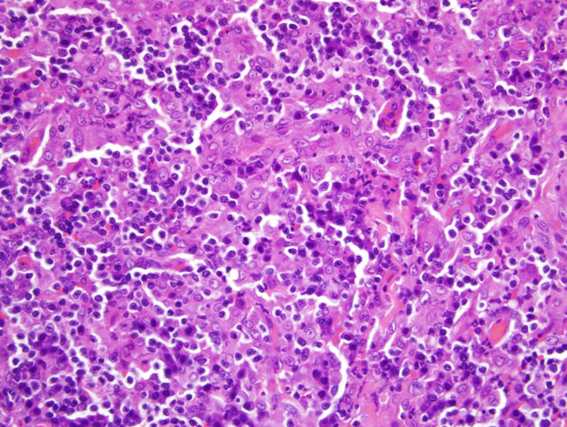
Histopathologic Description:
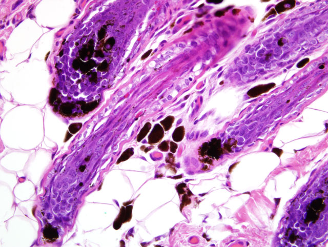
In the adjacent dermis, there is clumping of melanin within hair follicles with multiple melanophages in the surrounding dermis and mild periadnexal infiltrates of lymphocytes and plasma cells.
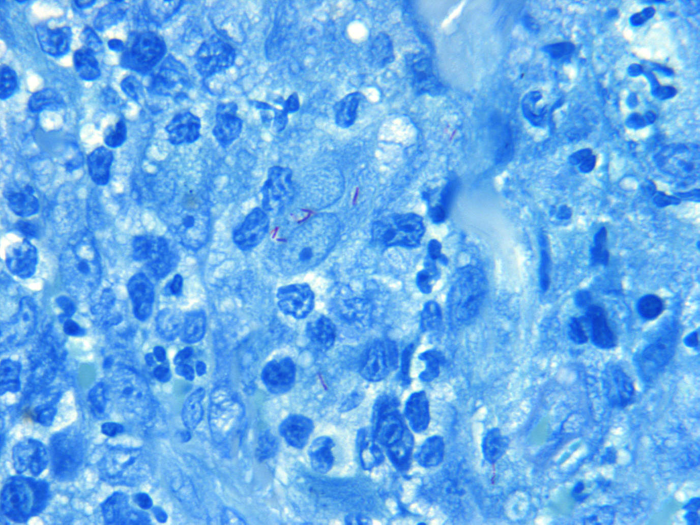
Ziehl-Neelson acid fast stain: Low numbers of acid-fast-positive bacilli, measuring 1-2 um in length, are within the cytoplasm of epithelioid macrophages.
Morphologic Diagnosis:
1. Haired skin (dorsal pinna): regional nodular granulomatous and lymphoplasmacytic dermatitis with focal ulcer and rare intrahistiocytic acid-fast bacilli (canine leproid granuloma syndrome).
2. Haired skin (dorsal pinna): follicular melanin clumping with dermal melanophages (possible color dilution alopecia).
Condition:
Contributor Comment:
On cytologic evaluation of fine-needle aspirates of the nodules, there are spindle-shaped macrophages with variable numbers of lymphocytes and plasma cells with fewer neutrophils and there are few to moderate numbers of negative-staining bacilli extracellularly or within macrophages.(1) On histopathology, there is dermal and/or subcutaneous granulomatous inflammation, with or without pyogranulomatous foci, and the inflammatory lesion consists mainly of epithelioid macrophages and neutrophils with lymphocytes, plasma cells, and rare multinucleated giant cells.(1,2,6) In Ziehl-Neelson acid-fast-stained specimens, there are very low to low numbers of intracellular bacilli in the majority of cases.(1,2,6)
The cause is a saprophytic, as yet unnamed and uncultivated, species of Mycobacterium.(1,2,4,6) Culture is unsuccessful, but with 16s rRNA PCR-based gene analysis on fresh and formalin-fixed, paraffin-embedded tissue, a proposed novel mycobacterial sequence has been identified.(4) The closest relatives of this agent are Mycobacterium tilburgii, M. simiae, and M. genavense.(4) There are significant molecular similarities between the organisms identified in the United States and those in Australia and New Zealand.(2) The suggested mode of transmission of the etiologic agent in canine leproid granuloma syndrome is thought to be percutaneous inoculation via wounds or biting insects.(2,6)
In the present case, the follicular melanin clumping with dermal melanophages is suggestive of concurrent color dilution in this dog. Color dilution alopecia is a hereditary skin disease in dogs with blue or fawn color-diluted coats and has been reported in the greyhound.(3) This disease is typically characterized by atrophy, distortion and abnormal melanin pigmentation of hair follicles with variable alopecia. Large amounts of clumped melanin pigment are within the hair follicle and within melanophages in the dermis around the base of hair follicles.(3) Some of the features of color dilution alopecia, such as the melanin clumping, may be seen in color-dilute dogs without alopecia and are not of pathologic significance.(3) Without enough supporting clinical history in this case, the contributors cannot determine whether this dog was color-diluted or fits the criteria for color dilution alopecia.
JPC Diagnosis:
1. Haired skin and subcutis: Dermatitis, pyogranulomatous, focally extensive, severe, with rare intrahistiocytic acid-fast bacilli.
2. Haired skin and subcutis: Follicular melanin clumping, multifocal, mild, with perifollicular pigmentary incontinence.
Conference Comment:
References:
2. Foley JE, Borjesson D, Gross TL, Rand C, Needham M, Poland A: Clinical, microscopic, and molecular aspects of canine leproid granuloma in the United States. Vet Pathol 39:234-239, 2002
3. Gross TL, Ihrke PJ, Walder EJ, Affolter VK: Skin Diseases of the Dog and Cat: Clinical and Histopathologic Diagnosis, 2nd ed., pp. 281-283, 518-522, 840-845, 866-872. Blackwell Publishing, Ames, IA, 2005
4. Hughes MS, James G, Ball N, Scally M, Malik R, Wigney DI, Martin P, Chen S, Mitchell D, Love DN: Identification by 16S rRNA gene analyses of a potential novel mycobacterial species as an etiologic agent of canine leproid granuloma syndrome. J Clin Microbiol 38:953-959, 2000
5. Lopez A: Respiratory system. In: Pathologic Basis of Veterinary Disease, eds. McGavin MD, Zachary JF, 4th ed., p. 520. Mosby Elsevier, St. Louis, MO, 2007
6. Malik R, Love DN, Wigney DI, Martin P: Mycobacterial nodular granulomas affecting the subcutis and skin of dogs (canine leproid granuloma syndrome). Aust Vet J 76:403-407, 1998
7. Malik R, Martin P, Wigney D, Swan D, Sattler PS, Cibilic D, Allen J, Mithcell DH, Chen SCA, Hughes MS, Love DN: Treatment of canine leproid granuloma syndrome: preliminary findings in seven dogs. Aust Vet J 79:30-36, 2001