Signalment:
Gross Description:
Histopathologic Description:
Morphologic Diagnosis:
Condition:
Contributor Comment:
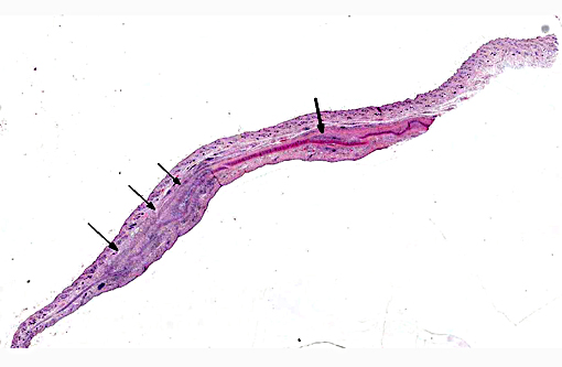
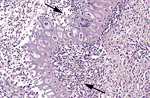
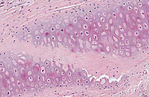
In cattle, one case report characterizes the lesions by lymphoplasmacytic infiltrates and presence of few macrophages within the cartilaginous plate of the pinna, which can be expanded by multiple basophilic cart-ilaginous nodules, vascularization and perivascular fibrosis. Chondrocytes in the center of the cartilaginous nodules may be swollen and found in clusters (proliferation). Rarely, low numbers of spindle cells surrounded by lacuna were present within these dense collagenous bundles (interpreted as early osseous metaplasia).6 In humans, histologically similar lesions may involve the pinnae, nose, trachea, joints, eyes and heart.(3)
JPC Diagnosis:
Conference Comment:
Feline relapsing polychondritis, more commonly known as auricular chondritis, is an uncommon condition in cats with no sex predilection affecting predominantly young to middle aged cats, although the lesion has been documented in older cats as well. The current published veterinary literature is unclear regarding whether additional cartilage tissues may be involved; although there has been speculation regarding the presence of joint, ocular lesions and cardiac involvement which is documented in the corresponding human condition as discussed above.(4) There is a case reported by Baba et al. which involved systemic joint and cartilage inflammation. In that case, the histologic ear lesions were similar to other cases reported in the literature. Additionally, costal cartilages were swollen, laryngeal cartilages thickened, and the articular cartilage of most peripheral joints eroded. There was also destruction of the subchondral bone, lymphocytic inflammation in the trachea and larynx and chondrolysis of the thyroid cartilage with associated inflammation. The costal cartilages were described as irregularly hypertrophic with bone formation and mixed inflammatory infiltration. Involvement of the respiratory tract and costal cartilage was not previously reported in cats. Uveitis was also reported in that case. Overall, the lesions described in Baba et al.s case report were more similar to human relapsing polychondritis than other cases of feline auricular chondritis previously reported. However, there were also discrepancies with the human condition, including the nature of the joint lesions, which were more consistent with, and diagnosed as chronic progressive arthritis. Additionally, the cat also had lymphoma, which was considered as a possible cause of the inflammatory lesions in the joints / cartilage.(2)
References:
1. Adissu HA, Baird JD, Wood GA. Case Report: A Case of Bilateral Auricular Chondritis in a Heifer. Case Reports in Veterinary Medicine. 2014; Article ID 929075. Hindawi Publishing. http://dx.doi.org/10.1155/2014/929075. Accessed June 2, 2015.
2. 6. Baba T, Shimizu A, Ohmuro T, Uchida N, et al. Auricular chondritis associated with systemic joint and cartilage inflammation in a cat. J Vet Med Sci. 2009;71(1):79-82.
3. Delmage DA, Kelly DF. Auricular chondritis in a cat. J Small Anim Pract. 2001;42(10):499-501.
4. Gerber B, Crottaz M, von Tschamer C, Scharer V. Feline relapsing polychondritis: two cases and a review of the literature. J Feline Med Surg. 2002;4(4):189-94.
5. Griffin, C. Dermatologic diseases of the auricle. 2006. International Veterinary Information Service; WSAVA lecture. www.ivis.org/proceedings/wsava/2006/lecture26/Griffin4.pdf - 2011-11-28. Accessed June 2, 2015.
6. Torres SMF. Miscellaneous Diseases of the Pinna. The Merck Veterinary Manual. Whitehouse Station, NJ: Merck and Co.;2009-2015. http://www.merckvetmanual.com/mvm/eye_and_ear/diseases_of_the_pinna/miscellaneous_diseases_of_the_pinna.html. Accessed June 2, 2015.