Signalment:
Gross Description:
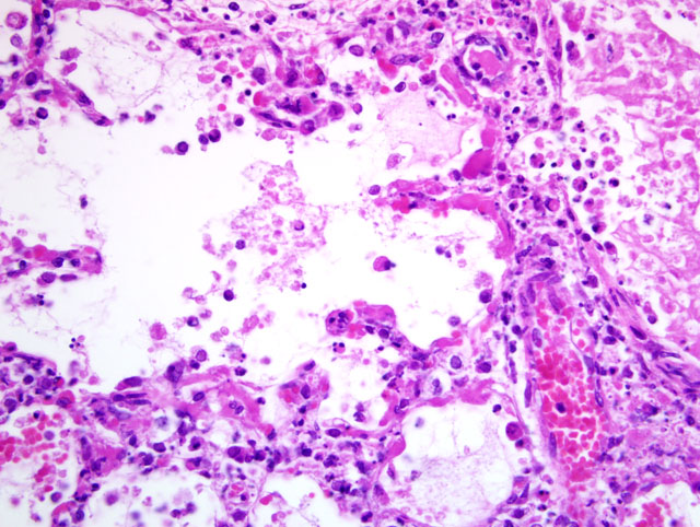
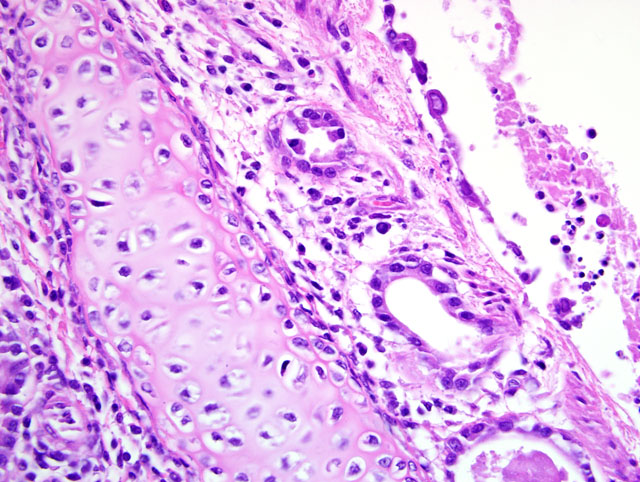
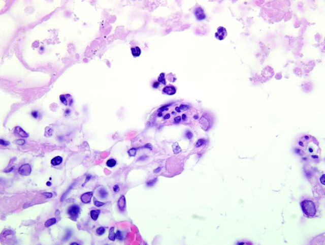
Histopathologic Description:
Morphologic Diagnosis:
Lab Results:
Condition:
Contributor Comment:
The classic disease associated with infection is 'feline rhinotracheitis,' most commonly manifested as upper respiratory disease with conjunctivitis in susceptible kittens. More recently, the virus has been implicated in cases of chronic conjunctivitis and periocular skin disease in adult cats.(5) Although abortion has been induced experimentally in pregnant queens by intravenous inoculation, it is not thought to occur with significant frequency as a natural disease.(2) Likewise, neonatal disease can be induced by exposure of kittens within the vaginal vault, but spontaneous cases of systemic disease in cats and kittens are rare.Â
Definitive diagnosis of herpesviral respiratory disease is often made clinically, but viral isolation from oropharyngeal swabs is possible if needed. The advent of PCR and in situ hybridization has made possible the implication of feline herpesvirus-1 in chronic keratoconjunctivitis and dermatitis as described above.(5)
Systemic disease characterized by interstitial pneumonia has been reported in kittens, (3) and in one case a seven-month-old cat with severe glossal ulcers was reported to have died of systemic illness characterized by multifocal hepatic necrosis with typical nuclear inclusions in hepatocytes.(4) In our case, a three-week-old Pixie-bob kitten had primary respiratory signs and at necropsy had lesions restricted to the lung. The diagnosis was suspected due to the presence of typical nuclear inclusions in pneumocytes and macrophages and was confirmed by viral isolation. Systemic herpesviral infections in kittens can be the result of immunosuppression, lack of protective antibodies or high pathogen load.(2) There are no reported genetic diseases of Pixie-bob cats that might predispose to severe viral infections. A possible scenario in this case would be stress-related recrudescence of FeHV-1 infection in the dam with transmission to a kitten with low protective antibodies. Chilling of a neonatal kitten to below body temperature may have allowed the virus to proliferate in systemic tissues.
JPC Diagnosis:
Conference Comment:
The contributor provided a succinct review of this entity. Conference participants also reviewed some less common clinical presentations of FeHV-1 infection, including ulcerative facial and nasal dermatitis, and stomatitis with eosinophilic infiltrates in cats and cheetahs.(2) For most participants, the differential diagnosis for necrotizing respiratory tract lesions in cats included feline calicivirus (FCV) infection, chlamydophilosis, and toxoplasmosis. When present, the characteristic herpesviral inclusions allow differentiation from FCV infection. However, herpesviral inclusions are only transient, and are usually absent after seven days post-infection. Herpesviruses are unique in that they elicit neutrophilic inflammation and fibrin exudation during respiratory infection, even in the absence of a secondary infection.(2) Feline calicivirus-induced pneumonia is generally interstitial in distribution, whereas FeHV-1 pneumonia is bronchointerstitial. Similarly, toxoplasmosis generally results in necrotizing multifocal to diffuse interstitial pneumonia, but the identification of intralesional protozoal cysts aids in making the diagnosis. Chlamydophila felis is an important cause of conjunctivitis in cats and a minor upper respiratory pathogen, but not an important cause of pulmonary disease.(1)
References:
2. Gaskell R, Dawson S, Radford A, Thiry Etienne: Feline herpesvirus. Vet Res 38:337-354, 2007
3. Love DN: Feline herpesvirus associated with interstitial pneumonia in a kitten. Vet Rec 89:178-181, 1971
4. Sheilds RP, Gaskin JM: Fatal, generalized feline viral rhinotracheitis in a young adult cat. J Am Vet Med Assoc 170:139-141, 1977
5. Suchy A, Bauder B, et al: Diagnosis of feline herpesvirus infection by immunohistochemistry, polymerase chain reaction and in situ hybridization. J Vet Diagn Invest 12:186-191, 2000