Signalment:
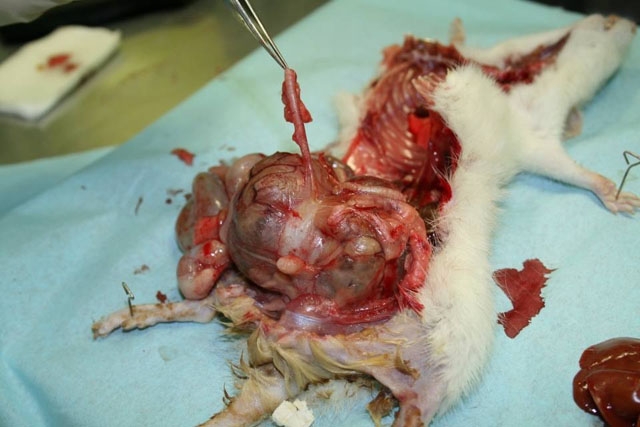
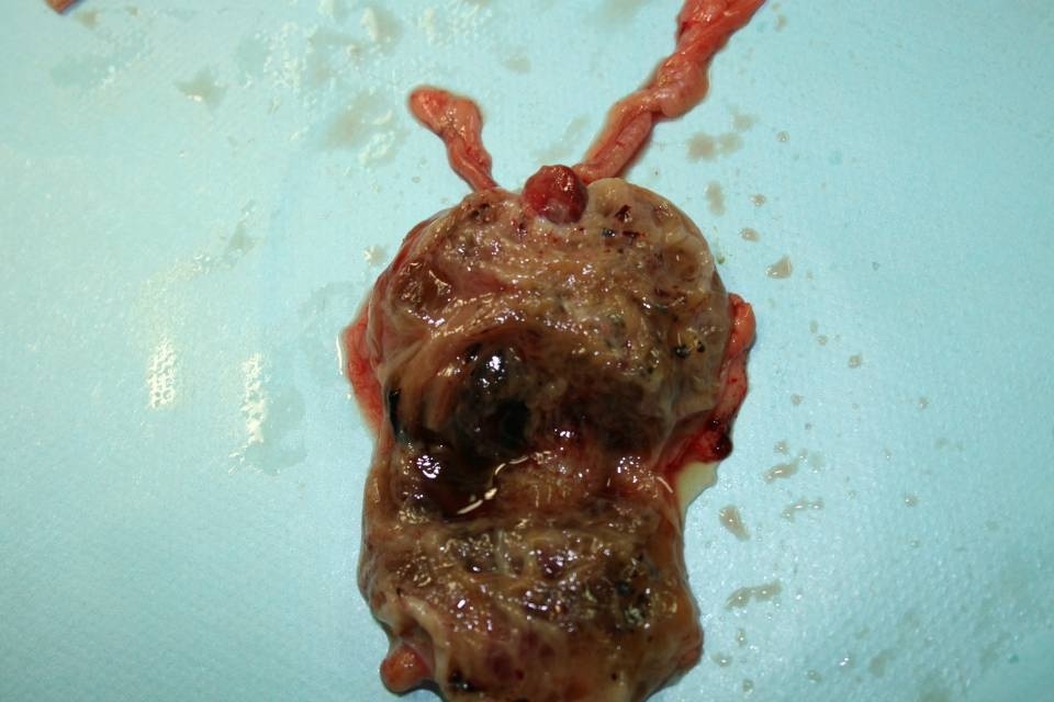
Gross Description:
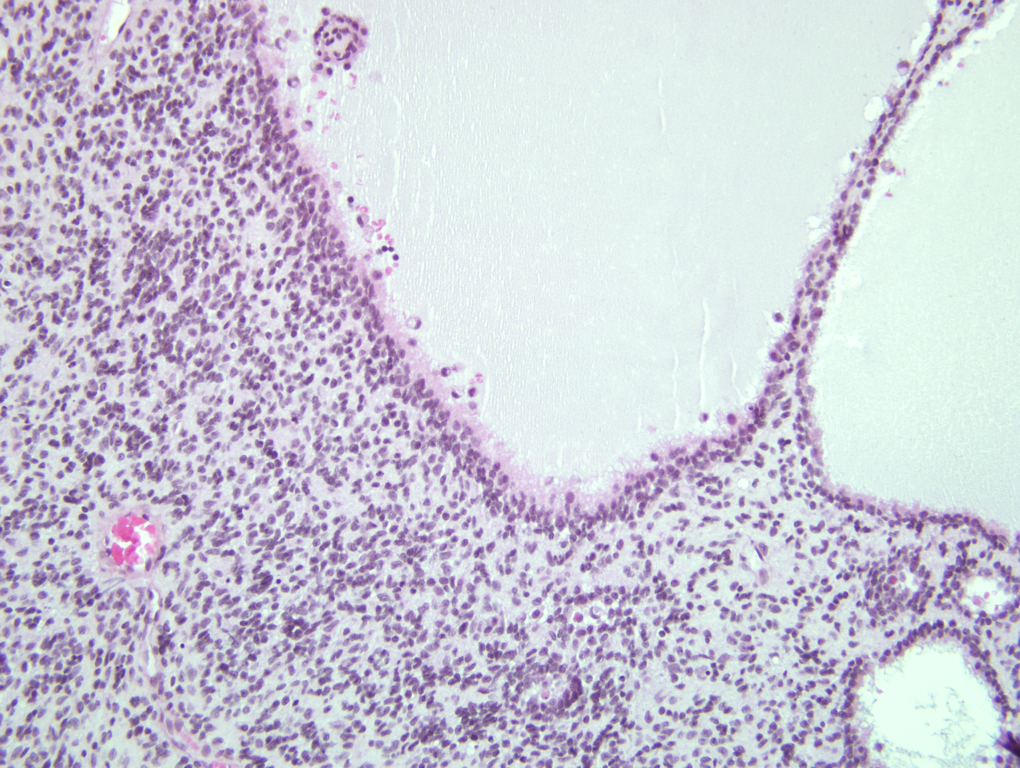
Histopathologic Description:
Uterine horn: The same neoplasm extends from the serosa to the lamina propria. The lumen is filled with pus.
Rectum and urethra: The same neoplasm invades the serosa and the tunica muscularis of the rectum.
Morphologic Diagnosis:
Lab Results:
Condition:
Contributor Comment:
The tumor in this case fulfills the typical morphological and immunohistochemical diagnostic criteria for uterine malignant schwannoma of the rat. The histologic appearance and demonstration of S-100 protein and GFAP by immunohistochemical procedures allow us to distinguish malignant schwannoma from the other mesenchymal neoplasms of the lower reproductive tract. Another criterion of distinction is the demonstration of basement membrane by electron microscopy.(1,7)
In human pathology, two patterns are often apparent in peripheral nerve sheath tumors: Antoni A and Antoni B patterns. Antoni A pattern is composed of compact spindle cells that usually have twisted nuclei, indistinct cytoplasmic borders, and occasionally clear intranuclear vacuole. They are arranged in short bundles or interlacing fascicles. In highly differentiated Antoni A pattern areas there may be nuclear palisading, whorling of the cells and Verocay bodies (two compact rows of well-aligned nuclei separated by fibrillar cell processes). Antoni B areas are less orderly and less cellular: they are composed of spindled or oval cells arranged haphazardly within the loosely textured matrix, which is punctuated by microcystic change, inflammatory cells, and delicate collagen fibers. As in Antoni B pattern areas the schwann cells possess increased number of lysosomes and myelin figures and the basal lamina is fragmented; the Antoni B pattern could be a degenerated Antoni A pattern.(5) Rat malignant schwannomas can have areas resembling these Antoni A and Antoni B patterns.(7) The tumor in this case is characterized by numerous pseudocysts and loosely packed cells reminiscent of Antoni B pattern.
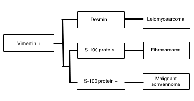
The differential diagnosis of malignant schwannoma of lower reproductive tract is detailed below:(9)
| Malignant Schwannoma | Cellular atypia Invasion Positive S-100 protein, vimentin immunoreactivity Negative immunostaining for desmin |
| Benign Schwannoma | No cellular atypia No invasion or distant metastasis |
| Fibrosarcoma | No basal lamina Negative S-100 protein immunoreactivity |
| Leiomyosarcoma | Eosinophilic blunt-ended nuclei Positive immunostaining for desmin |
Spontaneous peripheral nerve sheath tumors are rare in rats.(1) Malignant schwannomas can arise in the subcutis, stomach, heart, vagina and uterus.(1,7) In the endocardium, the differential diagnosis includes benign endocardial schwannomas, which occur more frequently. They are characterized by parallel arrays of cells, sometimes Antoni A-like pattern, and absence of prominent myocardial infiltration.(9) Endocardial schwann cell hyperplasia (neurofibromatosis) also occurs in the heart. It forms a thin layer beneath the endocardium and is well-demarcated from the myocardium; it is composed of less than 20 layers of cells.(9)
In both dogs and cats, malignant peripheral nerve sheath tumors are locally invasive and may have pulmonary metastases.(6) The primary sites of benign and malignant schwannomas are the brachial plexus, the lumbosacral plexus and the subcutis in the dog whereas in the cat the neoplasms arise in the nerve roots of the lower thoracic and upper lumbar cords segments.(6) In cattle, multifocal schwannomas are common in older animals and have a predilection for the autonomic nervous system, including the epicardial plexus, thoracic and cervical sympathetic ganglia, mediastinal nerve plexus, hepatic plexus, tongue, intercostal nerves, and brachial plexus.(6)
JPC Diagnosis:
Conference Comment:
Another differential diagnosis considered by few conference participants included endometrial stromal sarcoma. This uterine tumor often invades the myometrium, cervix and nearby abdominal organs. The neoplastic cells are spindled and arranged in streams, intersecting fascicles, or whorls on a fibrillar matrix. Although areas of necrosis, inflammation and hemorrhage may be seen, cysts or pseudocysts are not typically associated with endometrial stromal sarcomas. This tumor is typically immunohistochemically positive for S-100 protein and vimentin, and negative for desmin and actin. The spindle cell neoplasm in the case of this rat is immunopositive for glial fibrillary acidic protein (GFAP), which is most consistent with a tumor of peripheral nerve origin.(2)
The contributor provides an excellent synopsis of the comparative pathology of peripheral nerve sheath tumors.
References:
2. Dixon D, Leininger JR, Valerio MG, Johnson AN, Stabinski LG, Frith CH. Proliferative lesions of the ovary, uterus, vagina, cervix and oviduct in rats. In: Guides for Toxicologic Pathology; accessed at http://www.toxpath.org/nomen/index.htm, 11 August 2010. Washington D.C.: Society of Toxicologic Pathologists/American Registry of Pathology/The Armed Forces Institute of Pathology; 1999
3. do Amaral VF, Dal Lago EA, Kondo W, Souza LC, Francisco JC. Development of an experimental model of endometriosis in rats. Rev Col Bras Cir. 2009; 36(3):250-255.
4. Ellenson LH, Pirog EC. The female genital tract. In: Kumar V, Abbas AK, Fausto N, Aster JC, eds. Robbins and Cotran Pathologic Basis of Disease. 8th ed. Philadelphia, PA: Saunders Elsevier; 2010:1028-1029.Â
5. Enzinger FM, Weiss SW. Soft tissue tumors. St. Louis, MO: Mosby Press; 1995:829-837.
6. Koestner A, Higgins RJ. Tumors of the nervous system. In: Meuten DJ, ed. Tumors in Domestic Animals. 4th ed. Ames, IA: Iowa State Press; 2002:731-735.
7. Leininger JR, Jokinen MP. Oviduct, uterus and vagina. In: Boorman GA, Eustis SL, Elwell MR, Montgomery CA, MacKenzie WF, eds. Pathology of the Fischer Rat. San Diego, CA: San Diego Academic Press; 1990:455.Â
8. Mohr U. Soft tissue and musculoskeletal system. In: International Classification of Rodent Tumours, Part I - The Rat. Lyon, France: IARC Scientific Publications; 1992:34-36.Â
9. Mohr U. Central nervous system; Heart; Eye; Mesothelium. In: International Classification of Rodent Tumours, Part I - The Rat. Lyon, France: IARC Scientific Publications; 1992:34-37.