Signalment:
Gross Description:
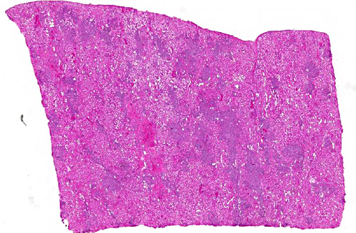
Histopathologic Description:
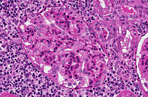
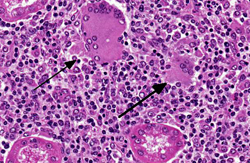
Granulomatous inflammation with multinucleated giant cells was also detected in the adrenal glands, pancreas, thyroid, heart, mammary glands, liver, uterus, skin, and lymph nodes. The condition in the heart, mammary glands, and skin was accompanied with eosinophilic infiltration.
Morphologic Diagnosis:
Lab Results:
| At clinical onset | At necropsy (9 days after the onset) | |
| Temperature (°C) | 38.1 | 39.3 |
| Heart rate (/min) | 84 | 90 |
| Respiration rate (/min) | 56 | 30 |
| White blood cells (/ml) | 9,100 | 12,300 |
| Packed cell volume (%) | 31 | 36 |
| Plasma protein level (g/dl) | 9.6 | 9.6 |
Flow cytometry analysis of the peripheral blood showed that the numbers of CD14+ monocytes (43.9%) and CD8 lymphocytes (19.1%) were significantly higher in the affected cows than in the normal lactating cows (CD14+ monocytes, 20.3 -¦ 1.6%; CD8 lymphocytes, 10.9 -¦ 1.3%). Further, the percentages of WC1 (3.5%) and IgM+ lymphocytes (6.7%) were significantly lower in the affected cows than in the normal lactating cows (WC1+ cells, 12.7 -¦ 2.5%; IgM+ cells, 15.9 -¦ 2.6%).
No pathogens were isolated from the carcass.
Condition:
Contributor Comment:
The granulomatous inflammation with the presence of multinucleated giant cells and the distribution of the lesions on the tissues are characteristics that are similar to those reported in hairy vetch toxicosis,(2,4,6) citrus pulp toxicosis,(3,7) and di-ureido isobutane (DUIB) toxicosis(1,5) in cattle. Various organs including the kidneys, heart, liver, spleen, adrenal glands, thyroid, lymph nodes, skin, and mammary gland are usually affected in these diseases.
Although the cause of the disease is an enigma, histopathological features suggest that a type 4 hypersensitivity reaction (key event) may play a role in the inflammatory reaction (pathogenesis).(6) We suggest that a plant constituent absorbed by the cows acts as an antigen that evokes a type 4 hypersensitivity reaction and granulomatous response. Alternatively, lectins may act as immunostimulants that directly stimulate T lymphocytes to initiate the inflammatory cytokine response that characterizes the disease. However, in this case, the diet that was fed to cows in the farm did not contain vetch, citrus pulp, or DUIB.
JPC Diagnosis:
Conference Comment:
The disease resembles a type 4 hypersensitivity reaction, although its pathogenesis is likely multifactorial as not all animals exposed develop lesions and lactating animals appear to be more susceptible.(4) The toxic principle was originally identified as prussic acid which is found in the seeds of Vicia villas Roth (hairy vetch),(3) a forage commonly cultivated as winter cover or in row crop rotations due its nitrogen-fixing ability. Other feed additives have been implicated (DUIB and citrus pulp), and this case serves as a reminder there are potentially other sources of a hapten or antigen capable of disease induction.
References:
1. Breukink HJ, Gruys E, Holzhauer C, Westenbroek AC: Pyrexia with dermatitis in dairy cows. Vet Rec 103: 221-222, 1978
2. Fighera RA, Barros CS: Systemic granulomatous disease in Brazilian cattle grazing pasture containing vetch (Vicia spp). Vet Hum Toxicol 46: 62-66, 2004
3. Hargis AM, Ginn PE. The integument. In: Zachary JF, McGavin MD, eds. Pathologic Basis of Veterinary Disease. 5th ed. St. Louis, MO: Elsevier Mosby; 2012:1018.
4. Iizuka A, Haritani M, Shiono M, Sato M, Fukuda O, Hagiwara A, Miyazaki S, Tanimura N, Kimura K, Nakazawa K, Kobayashi M, Takahashi T, Saito T, Fukai K: An outbreak of systemic granulomatous disease in cows with high milk yields. J Vet Med Sci 67: 693-699, 2005
5. Johnson B, Moore J, Woods LW, Galey FD: Systemic granulomatous disease in cattle in California associated with grazing hairy vetch (Vicia villosa). J Vet Diagn Invest 4: 360-362, 1992
6. Matsukawa K, Okada M, Kubo M: Pathomorphological findings of DUIB (1,1-Diureide isobutane) intoxication cases in dairy cattle. J Coll Dairying 10: 197-204, 1983 (in Japanese with English summary)
7. Panciera RJ, Mosier DA, Ritchey JW: Hairy vetch (Vicia villosa Roth) poisoning in cattle: update and experimental induction of disease. J Vet Diagn Invest 4: 318-325, 1992
8. Saunders GK, Blodgett DJ, Hutchins TA, Prater RM, Robertson JL, Friday PA, Scarratt WK: Suspected citrus pulp toxicosis in dairy cattle. J Vet Diagn Invest 12: 269-271, 2000