Signalment:
Adult female Friesian milk sheep (
Ovis ammon aries)While being kept for educational purposes during 2004 at the University of Veterinary Medicine Hannover, Clinic for Pigs, Small Ruminants, Forensic Medicine and Ambulatory Service, the sheep tested positive for infection with maedi-visna virus (MVV) and came in 2005 for further diagnostic reasons to the Friedrich-Loeffler-Institut, Federal Research Institute for Animal Health. The animal eventually developed in 2007 severe dyspnea and coughing, and became recumbent. It was then euthanized and submitted for necropsy.
Gross Description:
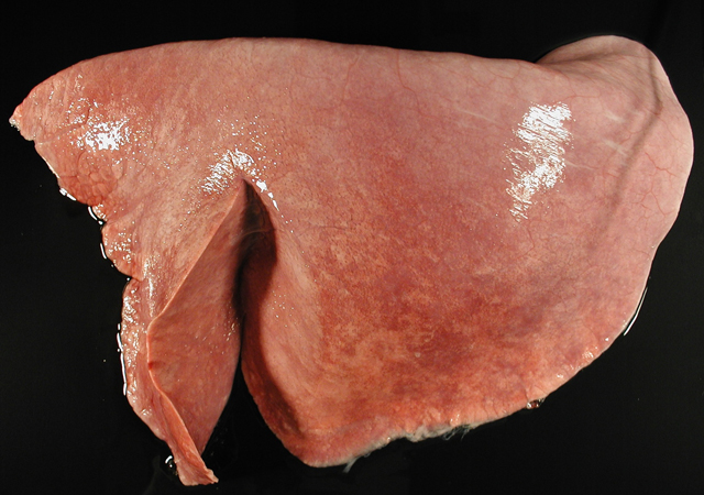
The lungs showed multifocal, subpleural and peribronchiolar nodular lesions up to 2 mm in diameter. These foci were grey and slightly elevated. Within the right cranial lobe there was a 3 x 2 x 2 cm sharply demarcated area of necrotizing and suppurative bronchopneumonia. The caudal mediastinal lymph nodes were enlarged and showed caseous necrosis and abscess formation.
Histopathologic Description:
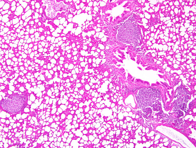
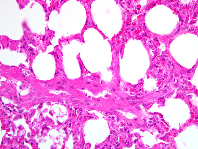
Lung: Multifocally, there are large, up to 1 mm in diameter, aggregates and sheaths of numerous lymphocytes and fewer macrophages in a follicular pattern (BALT hyperplasia) within the parenchyma or subpleural tissue, preferentially adjacent to bronchi, bronchioles and blood vessels. Diffusely, alveolar septa are slightly thickened by few infiltrating lymphocytes and macrophages and rare neutrophils. Around terminal bronchioles there are prominent bundles of smooth muscle cells (hypertrophy). Occasionally, in the alveolar lumina there are few detached pneumocytes.
Morphologic Diagnosis:
Lung: Pneumonia, bronchointerstitial, lymphocytic and histiocytic, multifocal, moderate, chronic with smooth muscle hypertrophy; etiology consistent with maedi-visna virus infection.
Lab Results:
Serological testing in 2004 by competitive ELISA revealed antibodies against MVV (more specific data not available). Bacteriological culture of lymph nodes revealed heavy growth of
Corynebacterium pseudotuberculosis admixed with
E. coli.
Condition:
Maedi-Visna Virus MVV, Ovine Progressive pneumonia
Contributor Comment:
Maedi is a persistent, chronic and progressive viral disease in sheep and goats that often results in lifelong infection. Cases have been documented in numerous countries worldwide, particularly continental Europe, the United Kingdom, Canada, the United States, Peru, Kenya, South Africa, Israel, India, Myanmar, and the southern regions of the former U.S.S.R. The word, maedi, is of Icelandic origin meaning, shortness of breath. However, as a disease found globally, it has been called by a variety of names-�-�Graaf-Reinet disease in South Africa, Zwoegerziekte in the Netherlands, La bouhite in France, and Montana, Marshs, or Ovine Progressive Pneumonia (OPP) as well as Lymphoid Interstitial Pneumonia (LIP) in the United States.(4) Maedi sometimes couples with visna, another slowly progressive disease causing meningoencephalitis in sheep and goats. Visna is the Icelandic word for wasting, which aptly characterizes a major clinical sign of the disease.
Maedi and visna are caused by the ovine maedi-visna virus complex (MVV). It is important to note, however, that the two diseases result from different strains of the virus.(6) MVV is a non-oncogenic retrovirus from the genus
Lentivirus in the
Retroviridae family. Retroviruses are spherical, enveloped virus particles with single-stranded, diploid, positive-sense RNA genomes. They possess a reverse transcriptase allowing them to integrate their genomes into the host cell DNA as a provirus. MVV is a small ruminant lentivirus (SRLV) exhibiting antigenic similarities to caprine arthritis encephalitis virus (CAEV). It can be subdivided into 5 groups (A-D) and further subtypes. MVV subtypes A1 and A2 seem to exclusively affect sheep, while A5, A7, B1, C, and D specifically affect goats. Subtypes A3, A4, A6 and B2 affect both sheep and goats.(5)
MVV is transmitted both horizontally and vertically. It can also be spread between species by direct contact. The primary mode of transmission has been via ingestion of colostrum or milk by the newborn.(5) Interestingly, there has been a higher risk of MVV infection when a newborn animal is bottle-fed colostrum from a seropositive ewe than when the newborn naturally suckles the same ewe.(3) Respiratory secretions constitute a secondary mode of transmission to ingestion of colostrum.(5)
Many sheep are seropositive for MVV, but few show clinical disease. The incubation period for the virus complex tends to be two to three years, possibly extending to eight.(6) Consequently, clinical signs will generally not appear in animals before they are two years old with the highest concentration of disease cases from five to ten years of age.(2) The primary manifestations of maedi are as follows: 1) a slow and progressively worsening dyspnea and 2) weight loss resulting in undernourishment, despite a normal appetite. With the appearance of clinical signs, maedi causes nearly 100% fatality.
On gross pathologic examination of maedi-diseased animals, lungs are heavier than normal and appear expanded. Rib impressions may also be apparent. The lungs will typically be mottled and gray. The cut surface is commonly dry and exudate cannot be extruded. Regional lymph nodes-�-�mediastinal and tracheobronchial-�-�are often enlarged.(4)
The general pathogenesis of MVV is not fully understood. Viral RNA and proviral DNA can be detected in a broad spectrum of cells, including dendritic cells, lymphocytes, plasma cells, endothelial cells, fibroblasts, adipocytes, microglial cells, pericytes, epithelial cells of bronchi, alveoli, mammary glands, thyroid follicles, choroid plexus, small intestine, renal tubules, and third eyelid. However, viral antigen has only been found in macrophages and monocytes, suggesting these cells may be the more exclusive replication sites for the virus complex.(1) The characteristic histopathologic finding for maedi is a pulmonary lymphocytic proliferation, predominantly of T cells.(4) Opportunistic bacterial infections can also occur, making a precise diagnosis of the disease sometimes difficult.Â
JPC Diagnosis:
Lung: Pneumonia, interstitial, lymphohistiocytic, diffuse, mild to moderate, with peribronchiolar lymphoid hyperplasia and smooth muscle hypertrophy.
Conference Comment:
The contributor provides a useful synopsis of this widespread entity. Lentiviral infections in small ruminants differ from those in felids and primates in that they do not result in immunosuppression. Progressive pneumonia is the most common manifestation of MVV infection in sheep; less frequent manifestations include encephalitis, arthritis, mastitis, and glomerulonephritis. Because it is closely related to MVV, CAEV not surprisingly produces a similar constellation of disease manifestations in goats (see WSC 2009-2010, Conference 1, case III), with several notable differences. Chief among these, arthritis in adults and neurologic disease in kids are far more common manifestations of CAEV in goats than is pneumonia. Additionally, CAEV pneumonia in goats is characterized by well-demarcated areas of type II pneumocyte hyperplasia, alveolar septal expansion by lymphocytes, and alveolar flooding with eosinophilic proteinaceous fluid. By contrast, type II pneumocyte hyperplasia is not a prominent feature of ovine progressive pneumonia, which is instead typified by interstitial pneumonia with the formation of lymphoid nodules with germinal centers, smooth muscle hypertrophy, and interstitial fibrosis.(2) In the present case, lymphoid proliferation and smooth muscle hypertrophy are the most striking lesions, although there is considerable slide variability with respect to severity, ranging from mild lymphocytic peribronchiolitis and perivasculitis to moderate lymphohistiocytic interstitial pneumonia as described above. There is infrequent type II pneumocyte hyperplasia, which participants attributed to local compression by immediately adjacent large lymphoid nodules.Â
References:
1. Brellou GD, Angelopoulou K, Poutahidis T, Vlemmas I: Detection of maedi-visna virus in the liver and heart of naturally infected sheep. J Comp Pathol
136:27-35, 2007
2. Caswell JL, Williams KJ: Respiratory system.Â
In: Jubb, Kennedy, and Palmers Pathology of Domestic Animals, ed. Maxie MG, 5th ed., vol. 2, pp. 618-620. Elsevier Saunders, Philadelphia, PA, 2007
3. Leginagoikoa I, Daltabuit-Test M, Alvarez V, Arranz J, Juste RA, Amorena B, de Andr+�-�s D, Luj+�-�n LL, Badiola JJ, Berriatua E: Horizontal maedi-visna virus (MVV) infection in adult dairy-sheep raised under varying MVV-infection pressures investigated by ELISA and PCR. Res Vet Sci
80:235-241, 2006
4. L³pez A. Respiratory System.Â
In: Pathologic Basis of Veterinary Diseases, ed. McGavin MD, Zachary JF, 4th ed., p. 533, Mosby Elsevier, St. Louis, MO, 2007
5. Pisoni G, Quasso A, Moroni P: Phylogenetic analysis of small-ruminant lentivirus subtype B1 in mixed flocks: evidence for natural transmission from goats to sheep. Virol
339:147-152, 2005
6. Zachary JF. Nervous System.Â
In: Pathologic Basis of Veterinary Diseases, ed. McGavin MD, Zachary JF, 4th ed., p. 936, Mosby Elsevier, St. Louis, MO, 2007