WSC 22-23
Conf 10
Case III:
Signalment:
5.5 year old, Female, New Zealand White Rabbit (Oryctolagus cuniculus)
History:
2 month history of "red urine". Initial urinalysis was within normal limits, with no RBCs detected. Radiographs revealed urinary bladder sludge, which was resolved after subcutaneous fluids. The rabbit was then reported for "red urine" again one month later. Another free-catch urinalysis was submitted which came back positive for red blood cells (too numerous to count) with high protein (100mg/dL) and 1+ cocci. CBC/Chemistry was within normal limits. The rabbit was euthanized at endpoint (for an ocular study) and submitted for necropsy.
Gross Pathology:
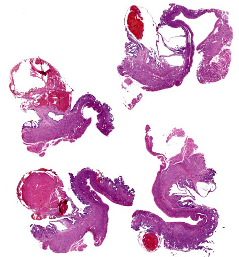
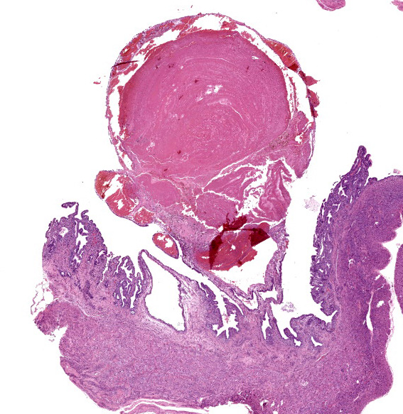
An adult female New Zealand White rabbit presents for necropsy several hours post euthanasia via intravenous euthasol. The carcass is in good post-mortem and adequate body condition with adequate subcutaneous and mildly increased visceral adipose stores. Externally, there is an intravenous catheter present in the left ear. There is mild focal yellow staining of the fur surrounding the perineum (urine). The bladder contains a mild amount of thick, moist, white to yellow sediment. The uterus is mildly enlarged, and there are multifocal raised nodules along the uterine horns bilaterally. On cut section, these nodules correspond to semi firm, papillary to polyploid masses that protrude into the uterine lumen, ranging from 1-3 cm in length and 0.5-1.5 cm in diameter. The endometrial surface between the polyploid masses contains numerous variably sized cysts and occasional foci of loosely adhered dark red gelatinous material (clotted blood) ranging from 1-2.5 cm in length. There are no other significant lesions.
Laboratory Results:
CBC/Chemistry: Within normal limits.
Urinalysis (free catch from pan): Red blood cells TNTC; protein 100 mg/dL; 1+ cocci
Microscopic Description:
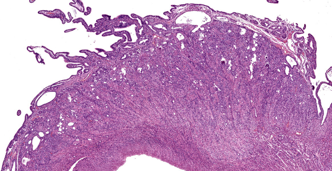
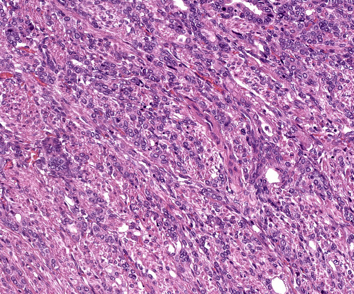
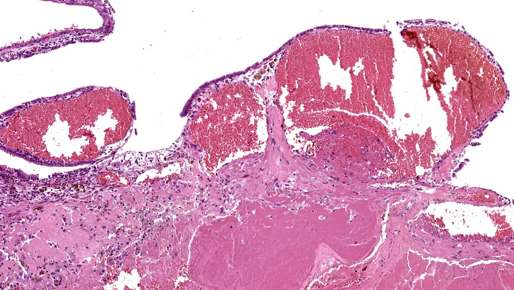
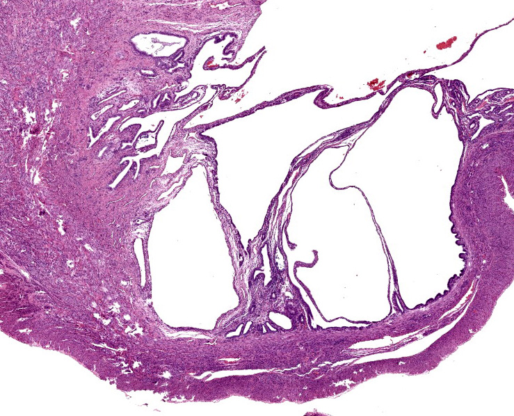
Uterus: Moderately expanding the endometrium and projecting into the uterine lumen are multifocal dilated veins which contain circular concentric lamellations of red blood cells and fibrin with scattered admixed leukocytes and plump fibroblasts (thrombus). Adjacent endometrium contains macrophages with intracellular brown pigment (hemosiderin, presumptive), mildly dilated blood vessels (congestion), and increased clear space (edema). Endometrial glands are mildly to moderately ectatic and hyperplastic, with attenuated or hyperplastic columnar epithelium occasionally thrown into papillary folds, and contain low numbers of histiocytes, erythrocytes, and basophilic acellular material. Dysplastic endometrial glands are also present with loss of nuclear polarity, angular glands projecting towards the myometrium, or intraluminal atypical endometrial cells. In other sections of uterus, multifocal uterine adenocarcinoma is present.
Contributor’s Morphologic Diagnoses:
Uterus: Endometrial venous aneurysms.
Uterus: Papillary adenocarcinoma, multicentric, with mild multifocal necrosis.
Uterus: Mild multifocal cystic endometrial hyperplasia.
Contributor’s Comment:
Hematuria in rabbits may be due to blood originating in the renal system or the reproductive system. Specific causes include neoplasia (uterine adenocarcinoma), uterine or bladder polyps, pyelonephritis or cystis, and urolithiasis.4,5 An additional cause of hematuria that appears to be unique to lagomorphs is endometrial venous aneurysms. This condition has been reported in multiple species of rabbit, including New Zealand White rabbits and a Holland Lop rabbit.4,7 The red urine in this case is reproductive in origin and associated with the uterine lesions. Histology of the lesion depicted on this slide was consistent with endometrial venous aneurysm. This rabbit also had multicentric uterine adenocarcinoma, which is not as well represented on this slide.
Grossly, endometrial aneurysms present as dark red, ovoid structures protruding into the uterine lumen from the endometrium, which corresponds microscopically to dilated venous structures containing blood or thrombi.5 Endometrial venous aneurysms are thought to be a congenital lesion and are characterized by localized venous dilation which may be fusiform or saccular in shape.7 In humans, congenital aneurysmal lesions of myometrial, cervical, and vaginal vessels but not endometrial veins have been reported.7 It has also been proposed that prolonged pseudopregnancy, which is common in rabbits and results in prolonged exposure to estrogen and progesterone, may also pay a role in increased vascularity or other vascular changes of the rabbit uterus.5 In a previous study, endometrial venous aneurysm was identified in 14/854 necropsy cases and 5/150 biopsy cases, with a median age of 32 months.3
For rabbits that present with hematuria secondary to endometrial venous aneurysms, ovariohysterectomy is recommended because of the risk of life-threatening hemorrhage.7 Ovariohysterectomy is curative in these cases.7
Contributing Institution:
University of Washington
Department of Comparative Medicine
Health Science Center
Seattle, WA 98105
JPC Diagnosis:
- Uterus: Uterine adenocarcinoma.
- Uterus: Endometrial venous aneurysms with thrombi.
- Uterus: Cystic endometrial hyperplasia, diffuse, moderate.
JPC Comment:
The contributor mentions several potential causes of hematuria originating from the reproductive tract in rabbits, including adenocarcinomas and endometrial hyperplasia. Several large-scale studies of neoplasms and uterine lesions in rabbits have demonstrated the relative frequency that these occur. According to several studies, uterine adenocarcinomas are the most frequently diagnosed uterine lesion in rabbits and the third most common neoplasm in rabbits in general, with only mammary carcinomas (20.2% of submissions) and trichoblastomas (18%) occurring with higher frequency in a study of 1238 rabbits.2,6,9 Most rabbits with uterine adenocarcinoma present with hematuria or serosanguinous vaginal discharge; anorexia is a less common clinical sign.9 The neoplasm is typically multicentric and nodular and may involve both uterine horns.1 The average age for diagnosis is 5-6 years of age, and the neoplasm tends to metastasize within 1-2 years to the lungs, liver, bone, or brain.9
Endometrial hyperplasia is the second most commonly diagnosed lesion of the rabbit uterus and outnumbers adenocarcinomas in some smaller scale studies.2,6,8,9,10 Endometrial hyperplasia occurred in 44% of 1928 rabbits with uterine lesions, and the most common clinical signs were similar to adenocarcinoma, with approximately half having hematuria or serosanguinous vaginal discharge and fewer having anorexia. The median age for endometrial hyperplasia is slightly younger, around 4 years.9,10 Additionally, it is common for adenocarcinoma to develop within areas of endometrial hyperplasia in rabbits, as was described in this case.2
In rabbits, hematuria must be differentiated from pigmented urine due to nonpathogenic crystals, porphyrins, or bilirubin by conducting a urinalysis, as was done in this case.1
References:
- Barthold SW, Griffey SM, Percy DH. Pathology of Laboratory Rodents and Rabbits. 4th Ames, IO: John Wiley and Sons, Inc. 2016: 256, 310, 320.
- Baum B. Not Just Uterine Adenocarcinoma – Neoplastic and Non-Neoplastic Masses in Domestic Pet Rabbits (Oryctolagus cuniculus): A Review. Vet Pathol. 2021; 58(5): 890-900.
- Bertram, CA, Müller K, and Klopfleisch R. Genital tract pathology in female pet rabbits (Oryctolagus cuniculus): a retrospective study of 854 necropsy examinations and 152 biopsy samples. J Comp Path 2018; 164: 17-26.
- Bray MV, Weir EC, Brownstein DG, and Delano ML. Endometrial venous aneurysms in three New Zealand White rabbits. Lab Anim Sci 1992; 42(4): 360-2.
- Diagnosis: Uterine Hemorrhage due to Endometrial Venous Aneurysms. Lab Anim 2003; 32(2): 24-25.
- Kunzel F, Grinniger P, Shibly S. Uterine Disorders in 50 Pet Rabbits. J Am Anim Hosp Assoc. 2015; 51(1): 8-14.
- Reimnitz L, Guzman D, Alex C, et al. Multiple Endometrial Venous Aneurysms in a Domestic Rabbit (Oryctolagus Cuniculus). J Exotic Pet Med 2017; 26: 230–237.
- Saito K, Nakanishi M, Hasegawa A. Uterine Disorders Diagnosed by Ventrotomy in 47 Rabbits. J Vet Me Sci. 2002; 64(6): 495-497.
- Settai K, Kondo H, Shibuya H. Assessment of reported uterine lesions diagnosed histologically after ovariohysterectomy in 1,928 pet rabbits (Oryctolagus cuniculus). J Am vet Med Assoc. 2020; 257(10): 1045-1050.
- Walter B, Poth T, Bohmer E, Braun J, Matis U. Uterine disorders in 59 rabbits. Vet Rec. 2010; 166: 230-233.