Signalment:
Gross Description:
Histopathologic Description:
Morphologic Diagnosis:
Condition:
Contributor Comment:
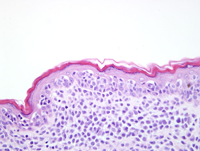
Two representative samples of the biopsy of the haired skin were submitted. In one section, there is marked epidermal ulceration with superficial bacterial colonization. The histopathologic lesions in each sample of the biopsy were characteristic of cutaneous epitheliotrophic lymphoma (abnormal lymphocytes localized in epidermis) (Fig. 4-1) .4, 5 This condition is thought to be analogous to mycosis fungoides (MF) in humans 1, except that in dogs, lymphocytic infiltrates consistently express CD3 and CD8, whereas in human cases, expression of CD 4 predominates.9 This condition is commonly observed in older dogs; however, its etiology is unknown .1,3
In this case, immunohistochemical stains were performed on paraffin-embedded sections with antibodies to the T-cell marker CD3 and the B-cell and plasma cell marker CD 79a. The lymphocytes that compose the population within the dermis and infiltrating the epidermis were strongly positive for CD3 antigen but were negative for CD 79a, indicating that this tumor had a T-lymphoid lineage consistent with immunohistochemical staining patterns previously reported in dogs.4
The disease progresses from erythematous patches and plaques to nodules and tumors, which are initially localized to the skin and mucous membranes but eventually spread to lymph nodes and metastasize .9 Due to the uncommon nature of the disease, diagnosis is often difficult. Differential diagnosis for skin lesions in canines include diseases like pemphigus vulgaris and discoid lupus or systemic lupus erythematosus lupus (immune-mediated diseases), histiocytoma, cutaneous histiocytosis and mastocytoma .5
This disease has a poor prognosis in the dog. Palliative treatment is often resorted to in order to prolong survival and maintain some reasonable quality of life for the animal .3,8 Anti-neoplastic drugs may induce temporary remission but are accompanied by side effects like contact dermatitis or cutaneous neoplasia.2,3,6 Radiotherapy is another option for treatment due to the radiosensitive nature of lymphoma cells .6
JPC Diagnosis:
Conference Comment:
The epitheliotropic form is further subclassified in the human literature into 4 forms, and although these forms do not generally correlate well clinically, they have been adapted by veterinary literature for histopathological classification:7
| Localized or generalized exfoliative erythroderma with scaling, alopecia, erosions or ulcerations without palpable masses Generalized form usually predominates in dogs and cats |
| Classical mycosis fungoides | Most common form seen in dogs Three stages (patch, plaque, and tumor) Patch and plaque stage usually seen simultaneously Progresses to tumor stage with spread to lymph nodes and other organs |
| dembl+�-�e form | Tumor formation without a previous patch or plaque stage |
| S+�-�zary syndrome | Circulating tumor cells in peripheral blood with epitheliotropic lymphoma and peripheral lymphadenopathy |
References:
2. Baker JL, Scott DW: Mycosis fungoides in two cats. J Am Anim Hosp Asso 25: 97-101, 1989
3. Beale KM, Bolon B: Canine cutaneous lymphosarcoma: Epitheliotropic and non- epitheliotropic, a retrospective study. In : Ihrke PJ, Mason IS, White SD, eds. Advances in Veterinary Dermatology., vol. 2, pp. 273-283. Oxford: Pergamon Pr, 1993 : 273-283, 1993
4. Bhang DH, Choi US, Kim MK, Choi EH, Kang MS, Hwang CY, Kim DY, Youn HY, Lee CW: Epitheliotropic cutaneous lymphoma (mycosis fungoides) in a dog. J Vet Sci 7: 97-99, 2006
5. Bouchard H: Epitheliotropic lymphoma in a dog. Can Vet J 41: 628-630, 2000
6. DeBoer DJ, Turrel JM, Moore PF: Mycosis fungoides in a dog : Demonstration of T-cell specificity and response to radiotherapy. J Am Anim Hosp Assoc 26: 566-572, 1990
7. Gross TL, Ihrke PJ, Walder EJ, Affolter VK: Lymphocytic tumors. In: Skin disease of the dog and the cat, 2nd ed., pp. 866-890, Blackwell Science, Ames, IA, 2005
8. Holloway KB, Flowers FP, Ramos-Caro FA: Therapeutic alternatives in cutaneous T-cell lymphoma. J Am Acad Dermatol 27: 367-378, 1992
9. Moore PF, Olivry T, Naydan D: Canine cutaneous epitheliotropic lymphoma (mycosis fungoides) is a proliferative disorder of CD8+ T cells. Am J Pathol 144: 421-429, 1994