Signalment:
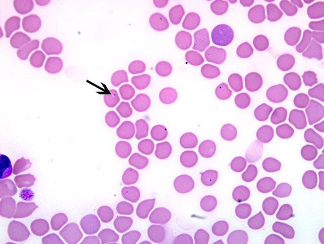
A blood smear was submitted for review.
Histopathologic Description:
Morphologic Diagnosis:
Lab Results:
CBC-
| DAY | 1 | 2 | 3(post transfusion) |
| Hematocrit (34-60%) | 18.5% | 15.8% | 30.2% |
| Platelet estimate | 128 K/μL | 48 K/μL | 128 K/μL |
| Segmented neutrophils (3,000-11,500/ μL) | 12,638/μL | NA | 2,686/ μL |
Chemistry no significant abnormalities
Prothrombin time 7.4 sec (5-12)
Partial Thromboplastin Time 7.8 sec (10-20)
Canine Direct Coombs test positive
Serology -
---- Babesia canis positive 1:320
---- Babesia gibsoni positive 1:640
---- Borrelia burgdorferi positive 1:640
PCR -
---- Babesia gibsoni positive for Asian genotype
Condition:
Contributor Comment:
The organisms are transmitted through the bite of an infected tick. Once inside the host, the organisms attach to the erythrocyte membrane and are endocytosed. While in the cytoplasm, the organisms undergo binary fission resulting in the formation of merozoites. Ticks become infected during feeding by ingesting merozoites. Transtadial and transovarial (B. gibsoni) transmission result in the formation of sporozoites within the ticks salivary glands. While feeding, saliva and sporozoites from the infected tick are passed into the hosts circulation. For transmission to occur, the tick must remain attached for 2-3 days.(2)
Infected dogs may present with a variety of clinical signs, ranging from hemolytic anemia to multiple-organ dysfunction syndrome (MODS). Subclinical infection is common in American pit bull terriers, which have a strong breed predilection for B. gibsoni. The predominant feature of babesiosis is hemolytic anemia. Thrombocytopenia is also quite common. The anemia is a result of both extra- and intravascular hemolysis. Parasitemia results in increased osmotic fragility, shortened erythrocyte life span, and erythrophagocytosis. The infected erythrocytes have parasite antigens on their surface, which induce opsonization by host antibodies and removal by the mononuclear-phagocyte system. The induction of serum hemolytic factors and production of antierythrocyte membrane antibodies with resulting damage from the secondary immune system also contribute to the pathogenesis of this disease. Other factors thought to be involved include oxidative damage, impaired hemoglobin function, as well as sludging and sequestration of infected erythrocytes.
Diagnosis may be made by identifying the parasite on a blood smear, serology (IFA and ELISA), or PCR which can detect low levels of parasitemia. Coinfection with other tick borne pathogens occurs relatively often as a single vector can harbor several infectious organisms.(1)
JPC Diagnosis:
Conference Comment:
Differential diagnoses for babesiosis include parasitic, immune-mediated, oxidative, and traumatic causes of hemolytic anemia.(3) Normally, infected animals present with anemia and thrombocytopenia. The anemia starts out as a normocytic, normochromic anemia and progresses to a macrocytic, hypochromic, regenerative anemia. Serum chemistries are generally within normal limits, with hypokalemia sometimes occurring in severely affected animals.(3)
Other intra-erythrocytic parasites of veterinary importance include:
| Species affected | Parasite |
| Dogs | Babesia canis, Babesia gibsoni |
| Cats | Babesia cati, Babesia felis |
| Cattle | Anaplasma marginale, Anaplasma centrale, Babesia bovis, Babesia bigemina, Theileria mutans, Theileria annulata |
| Sheep | Babesia ovis, Babesia motasi |
| Horses | Babesia equi, Babesia caballi |
| Deer, Elk | Theileria cervi |
| Birds | Hemoproteus spp., Leukocytozoon spp., Plasmodium spp. |
References:
2. Brockus CW, Andreasen CB: Erythrocytes. In: Duncan & Prasses Veterinary Laboratory Medicine, Clinical Pathology, eds. Latimer KS, Mahaffey EA, Prasse KW, 4th ed., pp. 19-21. Blackwell Publishing, Ames, IA, 2003
3. Taboada J, Lobetti R: Babesiosis. In: Infectious Diseases of the Dog and Cat, ed. Green CE, 3rd ed., pp 722-736. WB Saunders, St. Louis, MO. 2006