Signalment:
Gross Description:
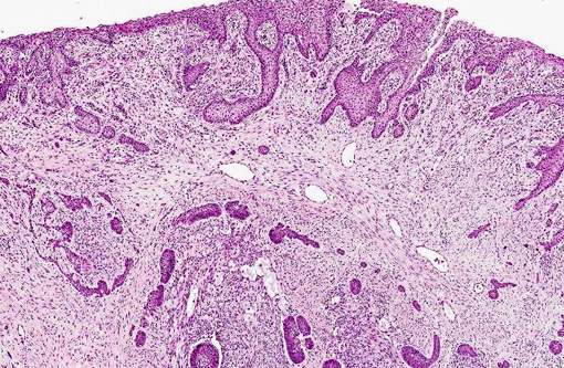
Histopathologic Description:
Morphologic Diagnosis:
Condition:
Contributor Comment:
Feline inductive odontogenic tumors (FIOT) are rare dental tumors specific to cats.(6,7) The neoplasm is the most common dental tumor of young kittens of either sex.(10) Feline inductive odontogenic tumor is described exclusively in cats under 3 years of age1 and is most often found on the rostral maxilla,(7,10) occasionally causing tooth loss or partial distortion. It has been confused with ameloblastoma in cats.(5) Unlike FIOT, which occurs in young cats, up to three years of age with most being younger than 18 months,(6) ameloblastomas occur in adult cats, chiefly over the age of six years and affects the maxilla and the mandible.(6)
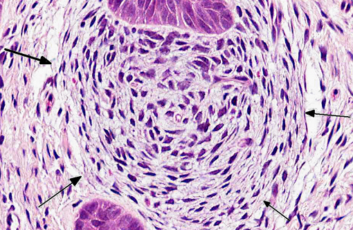
Feline inductive odontogenic tumor is a rare and interesting odontogenic neoplasm in which the odontogenic epithelium has inductive potential to form aggregated foci of dental pulp-like mesenchymal cells.(9) Its biological behavior is not well elucidated. The neoplasm is thought to have a benign behavior. Some studies indicate that Type IV collagen and laminin were constantly positive around the foci of epithelial cells, and Ki-67 positive indices were extremely low. These findings are consistent with the benign clinical presentation.(9) Although, it is a benign neoplastic mass histopathologically, feline inductive odontogenic tumor grows by expansion and can infiltrate the underlying bone and cause considerable local destruction.(1) Local recurrence has been reported in incompletely excised masses; however, metastasis does not appear to occur. This tumor differs microscopically from human ameloblastic fibromas in that it is not well-circumscribed but rather originates multifocally within the supporting connective tissue as characteristic, spherical condensations of fibroblastic connective tissue (ectomesenchyme) associated with islands of odontogenic epithelium.(6)
JPC Diagnosis:
Conference Comment:
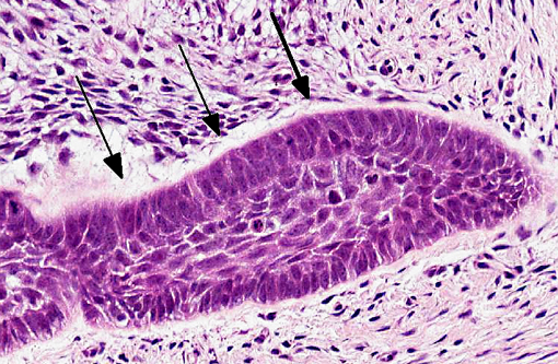
Odontogenic epithelium is characterized by 1) peripheral palisading of columnar epithelial cells, 2) apical hyperchromatic nuclei with basilar cytoplasmic clearing and 3) prominent intercellular bridging between internal epithelial cells (stellate reticulum).(2) Tumors of odontogenic epithelium are broadly divided into two categories: non-inductive tumors without odontogenic mesenchyme, including ameloblastoma, amyloid-producing odontogenic tumor and canine acanthomatous ameloblastoma, and inductive tumors with the presence odontogenic mesenchyme, including feline inductive odontogenic tumor, ameloblastic fibroma, complex odontoma and compound odontoma.(2,8,9,10)
Ameloblastoma, the least differentiated of the non-inductive odontogenic tumors, has been reported in dogs, cats, horses and humans. Ameloblastoma is classified as central (within the bone) or peripheral (within the gingival soft tissue) and is characterized by islands of poorly differentiated odontogenic epithelium, occasionally admixed with foci of metaplastic bone. Ameloblastic epithelial cells often undergo squamous differentiation and tumors are occasionally so heavily keratinized that it becomes difficult to differentiate ameloblastoma from squamous cell carcinoma. Central ameloblastomas are more common in animals.(2,8,10) The neoplastic islands of odontogenic epithelium in amyloid-producing odontogenic tumors (APOT) are separated by abundant waxy eosinophilic material (amyloid) which exhibits strong apple-green birefringence under polarized light when stained with Congo red. Recent studies have suggested the protein in APOT is not actually amyloid, but is derived from an ameoblastin-like peptide, or AAmel. Ameloblastin (formerly known as sheathlin) is an enamel matrix protein that is essential for enamel formation. Ameloblastin and another enamel protein, amelogenin, are both produced by ameloblasts during amelogenesis (see WSC 2012-13, conference 8, case 2).(2,4) Canine acanthomatous ameloblastoma is more locally aggressive than the other non-inductive odontogenic tumors and, like the ameloblastoma, is generally composed of interconnecting cords and sheets of odontogenic epithelium. It can be differentiated from ameloblastoma by an increased amount of stroma with abundant fibrillar collagen, regularly-positioned stellate mesenchymal cells, and regularly dispersed empty blood vessels, reminiscent of periodontal ligament connective tissue (see WSC 2012-13, conference 8, case 1). Cyst formation is common in acanthomatous ameloblastomas, however, in contrast to ameloblastomas, keratinization is rare.(2,8)
A comprehensive review of feline inductive odontogenic tumors is provided above by the contributor. Another inductive odontogenic tumor, ameloblastic fibroma, is composed of islands and cords of odontogenic epithelium on an abundant, collagen-poor fibrous stroma resembling dental pulp (see WSC 2011-12, conference 1, case 4). This is the most common odontogenic tumor in cattle, but has also been reported in young horses and dogs.(2,7) Complex/compound odontomas, the most differentiated odontogenic tumors, are composed of tooth-like structures known as denticles. Denticles contain enamel, dentin, cementum and pulp, arranged in a manner similar to a normal tooth; discrete islands of odontogenic epithelium are not present. These are reported in dogs, horses and cattle and can disrupt surrounding, normal teeth.(2)
References:
1. Beatty JA, Charles JA, Malik R, France MP, Hunt GB. Feline inductive odontogenic tumour in a Burmese cat. Aust Vet J. 200;78(7):452-455.
2. Brown CC, Baker DC, Barker IK. The alimentary system. In: Maxie MG, ed. Jubb, Kennedy and Palmers Pathology of Domestic Animals, 5th ed. Vol 2. Philadelphia, PA: Elsevier Saunders; 2007:5-7, 24-28.
3. Caceci T. Digestive System I: Oral Cavity & Associated Structures. VM8054: Veterinary Histology website. http://www.vetmed.vt.edu/education/Curriculum/VM8054/Labs/labtoc.htm. Accessed October 29, 2013.
4. Delaney MA, Singh K, Murphy CL, Solomon A, Nel S, Boy SC. Immunohistochemical and biochemical evidence of ameloblastic origin of feline amyloid-producing odontogenic tumors in cats. Vet Pathol. 2013;50(2):238-242.
5. Gardner DG. Ameloblastomas in cats: a critical evaluation of the literature and the addition of one example. J Oral Pathol Med. 1998;27(1):39-42.
6. Gardner DG, Dubielzig RR. Feline inductive odontogenic tumor (inductive fibroameloblastoma) a tumor unique to cats. Journal of Oral Pathology & Medicine. 1995;24:185190.
7. Head KW, Else RW, Dubielzig RR. Tumors of the alimentary tract. In Meuten DJ, ed. Tumors in Domestic Animals. 4th ed. Ames, IA: Iowa State Press; 2002:401-481.
8. Head KW, et al. Tumors of odontogenic epithelium without odontogenic mesenchyme. In: Tumors of the Alimentary System of Domestic Animals. Washington DC: AFIP and CL Davis DVM Foundation and WHO Collaborating Center for Worldwide Reference on Comparative Oncology; 2003:49-51.Â
9. Hiroki Sakai, Takashi Mori, Tsuneyoshi Iida, Yanai Tokuma, Kouji Maruo and Toshiaki Masegi. Immunohistochemical features of proliferative marker and basement membrane components of two feline inductive odontogenic tumours. Journal of Feline Medicine & Surgery. 2008;10(3):296-299.Â
10. Poulet FM, Valentine BA, Summers BA. A Survey of epithelial odontogenic tumors and cysts in dogs and cats. Vet Pathol. 1992;29:369-380.
11. Tutt C. Tooth development (odontogenesis). In: Small Animal Dentistry: A Manual of Techniques. 2006:1-32.