Signalment:
Gross Description:
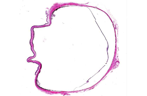
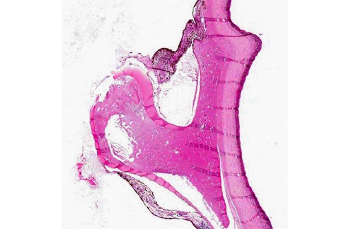
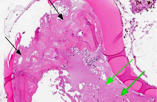
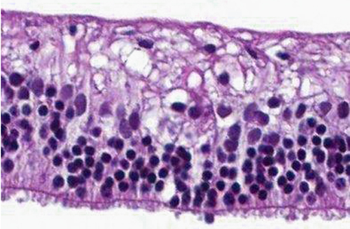
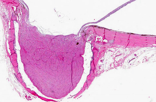
Histopathologic Description:
Morphologic Diagnosis:
Condition:
Contributor Comment:
If the zonular disinsertion is partial and the lens still rests in the patella fossa of the anterior vitreous, it is described as a posterior subluxation. If the zonular disinsertion is total, with migration of the lens posteriorly onto the floor of the vitreous, or anteriorly through the pupil, into the anterior chamber, the lens is described as luxated. Pathogenesis of sequelae involve the distortion of the visual optics, physical irritation by the displaced lens to retina, anterior uvea, and/or cornea, and pupillary block by the lens itself of the adherent vitreous with resultant secondary glaucoma. Luxated lenses become cataractous if they are not already, likely due to altered nutrition.
Primary luxation occurs in the terrier breeds; dogs are usually middle-aged and the condition is always bilateral although not necessarily concurrently so. The affected eye may present with iridodonesis, an aphakic crescent, and irritation manifested by redness, blepharospasm, and tearing if the lens is subluxated; IOP may be variable. Anterior luxation is characterized by visualization of the lens equator and obscuration of the pupil; IOP will usually be elevated and if it is not, it soon will be. Posterior luxations are diagnosed by observing a deep anterior chamber with a concavity to the iris surface, and the lens on the floor of the posterior segment; IOP is usually not elevated. Examine fellow eyes closely; iridodonesis may be present and dilation may reveal an aphakic crescent. Dilation may turn an innocuous posterior subluxation into an emergent anterior luxation. In cats, the majority of luxations occur secondary to chronic uveitis; secondary glaucoma is uncommon compared to the dog, related to the deep feline anterior chamber and the liquefaction of the vitreous that accompanies chronic inflammation.
Primary lens displacement has been documented to be heritable in many terrier breeds as well as Tibetan Terriers, Border Collies, and Shar-Peis. PLD is invariably a bilateral condition, albeit usually not simultaneous. When a patient presents with one eye affected, the fellow eye may vary in presentation from no apparent instability to early signs of instability such as iridodensis, phacodonesis, and vitreal herniation, to clear evidence of subluxation or complete luxation. The reported time to displacement in the fellow eye varies from days to years. Lens displacement often leads to vision- threatening complications, of which glaucoma is the most common and can occur regardless of lens position (anterior, posterior, or subluxated). Other reported complications include retinal detachment and uveitis.(4)
JPC Diagnosis:
Conference Comment:
Conference participants discussed the difficulty in determining primary versus secondary lens luxation from histology alone, concluding that similar lesions can be seen in both. As the contributor notes, primary luxation of the lens is due to rupture of the lens zonules, which function to keep the lens in its normal position between the ciliary processes. The zonular fibers are made of microfibrils (primarily the glycoproteins fibrillin-1 and fibrillin-2). Genetic abnormalities in genes coding for fibrillin-1 have been shown to cause lens displacement in humans and possibly in cattle.(6) A truncating gene mutation in ADAMTS17 on CFA03 has been found to be associated with the development of primary lens displacement in 17 dog breeds; however, its mode of inheritance and penetrance have yet to be fully elucidated.(2) Primary lens displacement in dogs appears to be affected by both dose and age (i.e. heterozygotes are affected later in life than homozygotes, and penetrance increases with age), which is consistent with an additive allelic model.(2) ADAMTS17 is a member of the secreted metalloproteinase family of proteins which bind extracellular matrix; it is believed to play a role in the formation of crystalline lens zonules and connective tissue. Mutations in ADAMTS17 cause abnormalities similar to Weill-Marchesani syndrome (WMS) in humans. WMS, which is associated with mutations in a related gene (ADAMTS10), as well as FBN1, is a rare connective tissue disorder in which patients develop eye and skeletal abnormalities.(5)
References:
2. Gharahkhani P, OLeary C, Duffy D, Bernays M, Kyaw-Tanner M. Primary lens luxation in Australian Tenterfield and Miniature Bull Terriers is due to an old ADAMTS17 mutation and is an additive trait. The Open Genomics Journal. 2012;5:7-13.Â
3. Gould D, Pettitt L, McLaughlin B, Holmes N, Forman O, Thomas A, Ahonen S, Lohi H, OLeary C, Sargan D, Mellersh C. ADAMTS17 mutation associated with primary lens luxation is widespread among breeds. Veterinary Ophthalmology. 2011;14, 6, 378384.
4. Gelatt K, MacKay E. Secondary glaucomas in the dog in North America. Veterinary Ophthalmology. 2004;7:245259.Â
5. Morales J, Al-Sharif L, Khalil DS, Shinwari JMA, Bavi P, Al-Mahrouqi RA, et al. Homozygous mutations in ADAMTS10 and ADAMTS17 cause lenticular myopia, ectopia lentis, glaucoma, spherophakia, and short stature. Am J Hum Genet. 2009;85(5):558568.
6. Morris RA, Dubielzig RR. Light-microscopy evaluation of zonular fiber morphology in dogs with glaucoma: secondary to lens displacement. Vet Ophthalmol. 2005;8(2)81-44.Â
7. Sargan DR, Withers D, Pettiite I, Squire M, Gould DJ, Mellersh CS. Mapping the mutation causing lens luxation in several terrier breeds. Journal of Heredity. 2007:98(5):534538.