Signalment:
Yearling, male, Harbor seal (
Phoca vitulina).This animal was found dead on a beach in the Pacific Northwest.
Gross Description:
The animal was emaciated and featured corneal abrasions and numerous nasal mites.
Histopathologic Description:
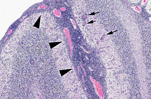
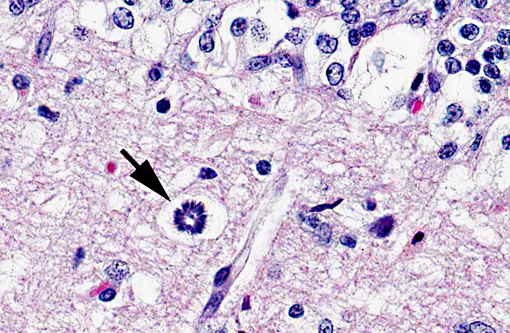
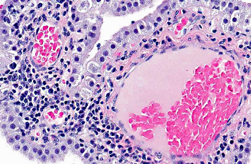
Throughout the cerebellum, expanding the meninges, extending focally to segmentally throughout the molecular layer and expanding and disrupting the molecular and granular layers of numerous folia, there is multifocal to coalescing accumulations of lymphocytes and histiocytes with fewer plasma cells. In more severely affected areas, there is multifocal edema fluid with neovascularization, reactive endothelia, lymphoplasmacytic perivascular cuffing and edema. Randomly interspersed within the inflammatory infiltrate are numerous protozoa with multifocal karyorrhectic and pyknotic debris.
Morphologic Diagnosis:
Brain: Meningoencephalitis, severe, multifocally extensive, necrotizing, lymphohistiocytic, with numerous intralesional protozoa.
Lab Results:
PCR of pooled tissues proved negative for canine distemper virus and influenza virus and positive for
Apicomplexa. No bacteria were recovered from the lung, lymph node, brain or spleen and there was heavy growth of
Escherichia coli from the small intestine. Trace mineral and vitamin A analysis results of the liver proved within in house reference limits.
Condition:
Sarcocystis neurona
Contributor Comment:
Since 2000, increasing numbers of marine mammals have stranded within the Pacific Northwest with protozoal encephalitis. In the initial stages of this phenomenon,
Toxoplasma gondii was the primary pathogen, however, more recently,
Sarcocystis neurona has emerged as the dominant etiologic agent. Based on review of brain pathology, dual infections appear to have potentiated the pathogenicity of both parasites. Malnutrition, contaminant loads and other environmental stressors may have also potentiated the pathology of infections. The increase in
S. neurona infections is attributed to the extension of opossums north, into Washington State and British Columbia.
JPC Diagnosis:
Cerebellum and brain stem: Encephalitis, necrotizing, multifocal to coalescing, severe, with diffuse lymphohistiocytic and neutrophilic meningitis, choroid plexitis, and numerous intracellular schizonts.
Conference Comment:
Conference participants discussed the differential diagnosis for this case, and favored
Sarcocystis neuron; however, since mature schizonts and merozoites of
Sarcocystis are difficult to distinguish from
Toxoplasma, we consulted with Dr. J.P. Dubey. Immunohistochemical staining with antibodies against
Sarcocystis neurona performed by Dr. Dubey resulted in strong positive immunoreactivity in numerous schizonts. Furthermore, the diagnosis was confirmed by Dr. Dubey based on the morphology of the immature schizonts, as
S. neurona immature schizonts are morphologically unique in that their nucleus becomes multilobed, eventually giving rise to many merozoites, and often leaving a residual body. (J.P. Dubey, personal communication).
S. neurona is an apicomplexan, best known for causing equine protozoal myeloencephalitis (EPM), a severe neurologic disease in horses.(1) Opossums spread the disease by shedding sporocysts into the environment.(1) In addition to horses and sea mammals (sea otters, Pacific harbor seals), other animals including cats, striped skunks, and nine-banded armadillos can be infected with
S. neurona; however, in these species, the sarcocysts tend to develop in muscle rather than the central nervous system.(1,2) Raccoons have also been reported to have myocarditis and encephalitis due to infection with
S. neurona.(1)
References:
1. Bowman DD. Protozoans. In:
Georgis Parasitology for Veterinarians. 9th ed. St. Louis, MO: Saunders Elsevier; 2009:104-105.
2. Dubey JP, et al.
Toxoplasma gondii, Neospora caninum, Sarcocystis neurona, and
Sarcocystis canis-like infections in marine mammals.
Vet Parasitol. 2003;116(4):275-96.