CASE III:
Signalment:
Angus feeder calf (Bos Taurus) less than one year of age. Unknown gender.
History:
Acute onset of severe respiratory distress with cough and expiratory noise. The calf was afrebrile (102.6 °F). When the calf was removed from its pen to administer treatment, it died in the chute.
Gross Pathology:
No gross lesions were reported.
Laboratory Results:
PCR on lung tissue was negative for Pasteurella multocida, Histophilus somni, Mannheimia haemolytica, Mycoplasma bovis, Bovine herpesvirus 1, Bovine viral diarrhea virus, Bovine coronavirus, and Bovine respiratory syncytial virus.
Aerobic culture of lung yielded no bacterial pathogens.
Microscopic Description:
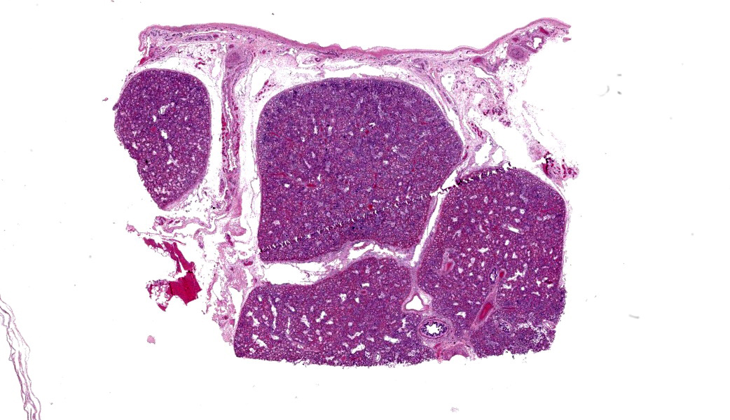
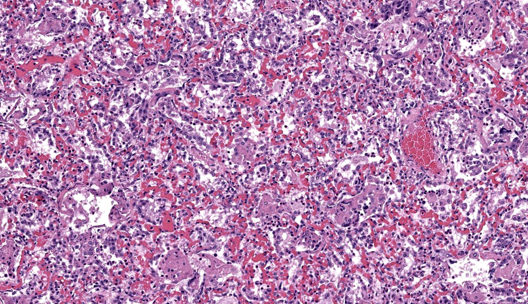
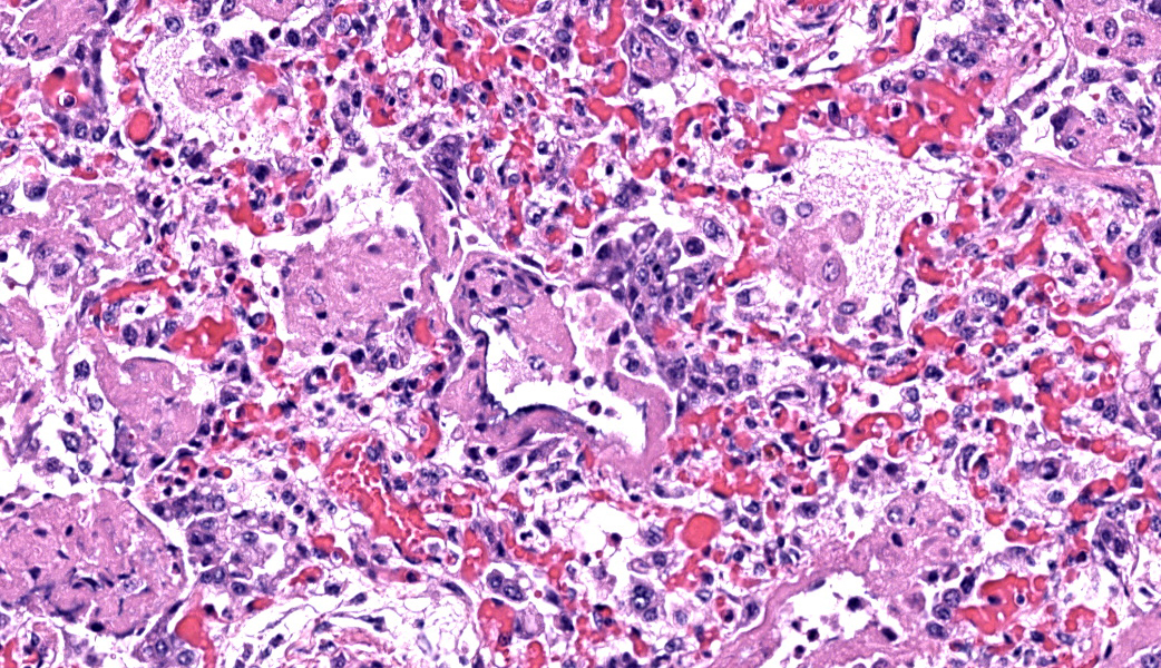
Lung: Sections of lung display a severe diffuse proliferative and exudative inflammatory process. Alveolar spaces contain variable amounts of homogenous eosinophilic fluid (edema fluid) mixed with fibrin and leukocytes. There is diffuse type II pneumocyte hyperplasia. Alveoli are lined by hyaline membranes (variability in the severity of hyaline membranes is observed among the sections submitted). Bronchi and bronchioles in some locations appear within normal limits whereas in other regions they are lined by attenuated epithelial cells and contain edema mixed with macrophages and sloughed epithelial cells within their lumina. There is pleural, peribronchiolar, and interlobular edema. Alveolar septa are thickened and severely congested.
Contributor Morphologic Diagnosis:
Lung; pneumonia, interstitial, histiocytic, subacute, diffuse, severe, with hyaline membrane formation, type II pneumocyte hyperplasia, and edema
Contributor Comment:
Atypical interstitial pneumonia (AIP) represents a sporadic disease of cattle that has traditionally been known by a number of names including "fog fever", "dust pneumonia", and "acute bovine pulmonary emphysema and edema", among others.5,7 A number of causes have been proposed in the literature including ingestion of compounds toxic to pneumocytes, poor air quality, inhalation of foreign material, as well as parasites and viruses, most notably BRSV.4
The metabolic activation and accumulation of 3-methyl indole (3-MI), a metabolite of tryptophan is the most widely accepted cause and is traditionally seen in cattle moved to lush green pastures in the early fall. Animals affected with AIP have been shown to have significantly higher plasma levels of 3-MI compared to unaffected animals.1 Additionally, a syndrome mimicking AIP was experimentally reproduced in cattle and goats after the administration of 3-MI.3
Within the rumen, L-tryptophan is converted first to indoleacetic acid and subsequently to 3-MI by Lactobacillus spp. of bacteria.2 Within the lung, nonciliated epithelial cells called Club cells (formerly called Clara cells) further metabolize 3-MI resulting in a compound that is toxic to pneumocytes and endothelial cells.4 The resulting vascular and epithelial cell damage results in large amounts of high protein content edema fluid that polymerizes forming the hallmark histologic feature of hyaline membranes. The lining of alveolar walls with hyaline membranes decreases oxygen exchange efficiency leading to the severe respiratory distress and ultimately death in these animals.
Further complicating the etiologic component behind AIP is the fact that the disease also occurs in feedlot animals who are not fed lush grasses but instead a more concentrated ration. Potential AIP causes or inciting factors unique to feedlot environments that have been proposed are discussed in a recent review and include melengestrol acetate (MGA), BRSV infection, bacterial respiratory disease, heat, and dusty conditions.9
Response to treatment for AIP is traditionally very poor.4 In the contributor's experience, feedlot cases of AIP tend to happen in fat cattle (most often heifers) at the end of the feeding period during the late summer months with high daytime temperatures. Experiencing sudden deaths due to AIP the week, or even the day of shipment to slaughter is not uncommon, making the economic losses even greater on a per case basis. The contributor has observed numerous cases with a history like this case where the stress of movement in these respiratory compromised animals proves too much and they often die on the way to, or within the chute when treatment is attempted.
Contributing Institution:
University of Nebraska -- Lincoln Nebraska Veterinary Diagnostic Center Lincoln, NE https://vbms.unl.edu/nvdlsJPC Morphologic Diagnosis:
Lung: Pneumonia, interstitial, necrotizing, subacute, diffuse, severe, with hyaline membranes, type II pneumocyte hyperplasia, and interlobular and pleural emphysema and edema.
JPC Comment:
This case illustrates a classic picture of acute interstitial pneumonia (AIP) in cattle. This condition has been historically known by a few different names, including "atypical interstitial pneumonia", "acute bovine pulmonary emphysema and edema" (ABPEE) or "fog fever." The names "fog fever" and ABPEE are still widely used clinically. Participants were reminded that this syndrome fits under the clinical umbrella of acute respiratory distress syndrome (ARDS), whereas pathologically it is best described as diffuse alveolar damage (DAD). It is important to note that DAD is a pattern rather than a single disease entity. Dr. Brown discussed with participants the five major causes of DAD in cattle that are worth having as a mental list: Lush pasture/L tryptophan (3 MI ("fog fever")), moldy sweet potatoes (4-ipomeanol), perilla mint toxicity, high oxygen exposure ("oxygen toxicity"), and radiation/certain chemotherapeutics.
On histology, the lung showed the expected constellation of hyaline membranes, type II pneumocyte hyperplasia, and interlobular and subpleural emphysema that are consistent with DAD. The group focused temporarily on the interlobular septa, where the clear spaces were debated as edema versus emphysema. Several participants emphasized that, in this context, the expansion of the interstitium and septa are far more likely to be emphysema from ruptured alveoli rather than just edema, especially when accompanied by the clear space dissecting along septa and into the pleura. This correlates well with the most reported gross findings for AIP in cattle, which include interlobular emphysema, a "checkerboard" appearance to the lungs of alternating firm, dark red areas and fluffy, pink areas, and overinflated lobes. 8
The pathogenesis discussion centered on the classic pasture-associated form of AIP, which is covered by the contributor in their comment. The resulting vascular and epithelial injury leads to protein-rich edema, hyaline membrane formation, and interstitial emphysema, all of which were seen in this case. The feedlot form of AIP received special attention because, as noted by the contributor, its pathogenesis differs. Unlike the well-characterized pasture-associated 3-MI form, the etiology of AIP in feedlot cattle remains unclear. While 3-MI toxicity is central to the disease in pastured cattle, evidence for the same mechanism in feedlot animals is lacking.6 Other factors mentioned by the contributor, including dietary composition, ruminal microbiome, concurrent respiratory disease, heat, dust, MGA administration in heifers, and management stressors, are all under consideration in the literature and were discussed in conference.6,8
Finally, conference goers addressed the chronicity (or lack thereof) of this case, which puzzled many participants. One participant remarked that, in subacute DAD, one expects fibrin and hyaline membranes whereas, in more chronic stages, type II pneumocyte hyperplasia predominates as the lung attempts repair. It is unusual, but not impossible, to see prominent hyaline membranes and marked type II pneumocyte hyperplasia together. When they do coexist, it likely reflects a lesion in transition, with ongoing injury superimposed on early repair.8 Both lesions together have been reported in cases of AIP in cattle.8 In fact, in a recent review, the most consistent histologic features in cases of AIP included hyaline membranes, type II pneumocyte hyperplasia, obliterative bronchiolitis, and interlobular emphysema.8 This nuance helped reconcile the features seen histologically with the clinical history of acute, severe respiratory distress.
References:
- Ayroud M, Popp JD, VanderKop MA, et al. Characterization of acute interstitial pneumonia in cattle in southern Alberta feedyards. Can Vet J. 2000;41(7):547-554.
- Carlson JR, Breeze RG. Ruminal metabolism of plant toxins with emphasis on in-dolic compounds. J Anim Sci.1984;58(4):1040-9.
- Carlson J, Yokoyama M, Dickinson, EO. Induction of Pulmonary Edema and Emphysema in Cattle and Goats with 3-Methylindole. Science. 1972;176:298-299.
- Doster AR. Bovine atypical interstitial pneumonia. Vet Clin North Am Food Anim Pract. 2010;26(2):395-407.
- Doster AR, Mitchell FE, Farrell RL, Wilson BJ. Effects of 4-ipomeanol, a product from mold-damaged sweet potatoes, on the bovine lung. Vet Pathol. 1978;15(3):367-75.
- Haydock LAJ, Fenton RK, Sergejewich L, Squires EJ, Caswell JL. Acute interstitial pneumonia and the biology of 3-methylindole in feedlot cattle. Anim Health Res Rev. 2022 Jun;23(1):72-81.
- Maxie MG, Jubb KVF. Jubb, Kennedy, and Palmer's Pathology of Domestic Animals. 2016:574.
- Schmidt PH, White BJ, Woolums AR, Larson RL. Scoping review of clinical signs, etiologies, pathology findings, risk factors, and treatments of acute interstitial pneumonia in North American feedlot cattle. JVDI. 2026:10406387261419913.
- Woolums AR. Feedlot Acute Interstitial Pneumonia. Vet Clin North Am Food Anim Pract. 2015;31(3):381-9.