Conference 24
CASE IV:
Signalment:
3-year-old intact male Rocky Mountain bighorn sheep (Ovis canadensis canadensis).
History:
One of a group of 17 rams from a provincial park. Over a period of ~1 month several rams were observed coughing and 5 were found dead. Several animals within this group were collared allowing monitoring of movements and recovery of carcasses. Movement monitoring revealed recent proximity to a flock of domestic sheep. At the time of euthanasia, this individual appeared lethargic and had been observed coughing. This animal was euthanized by gunshot to the head and submitted for necropsy to allow for further evaluation of respiratory disease for herd management. The carcass was stored outside in -20oC temperatures for ~12 hours prior to necropsy examination.
Gross Pathology:
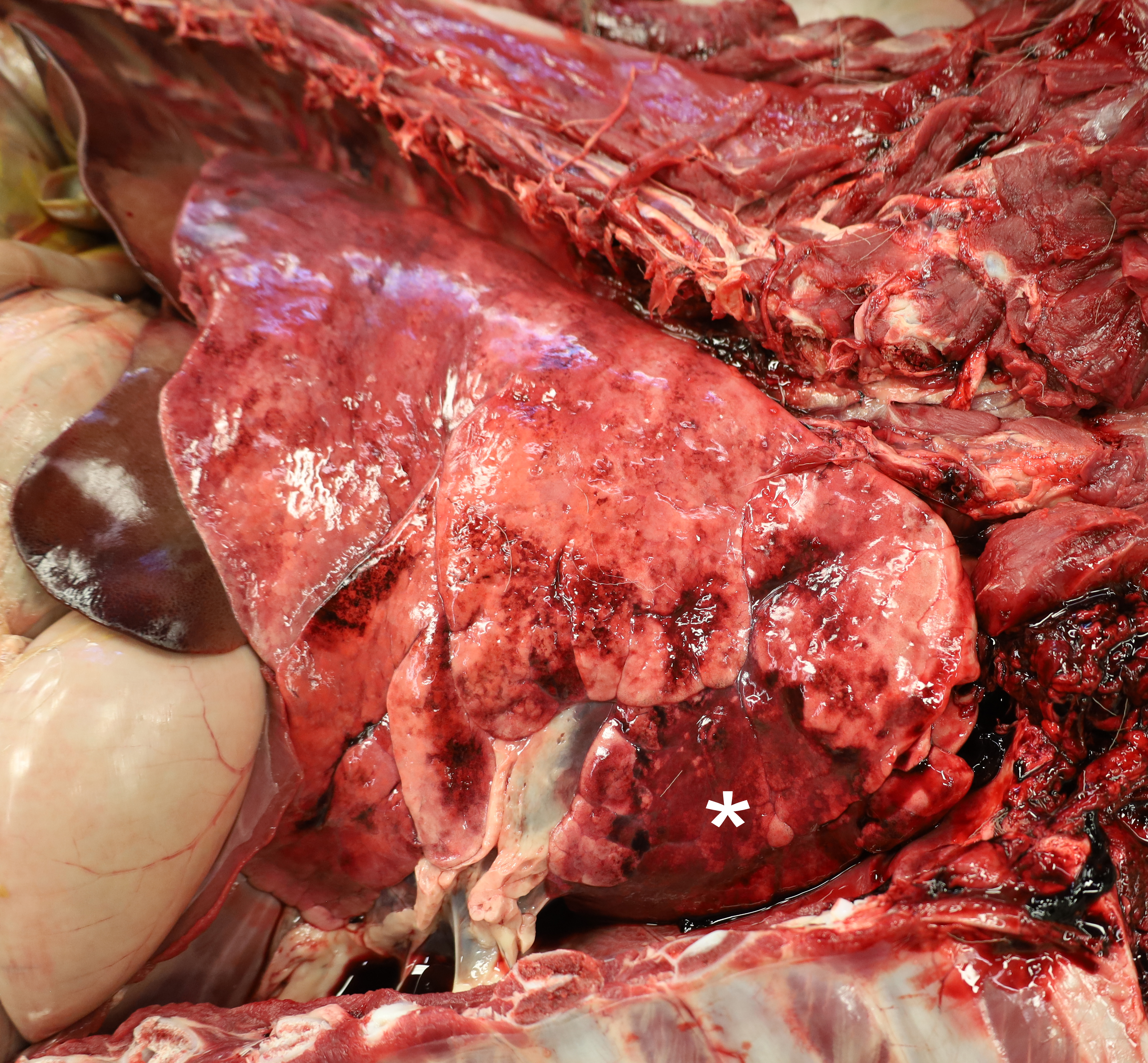
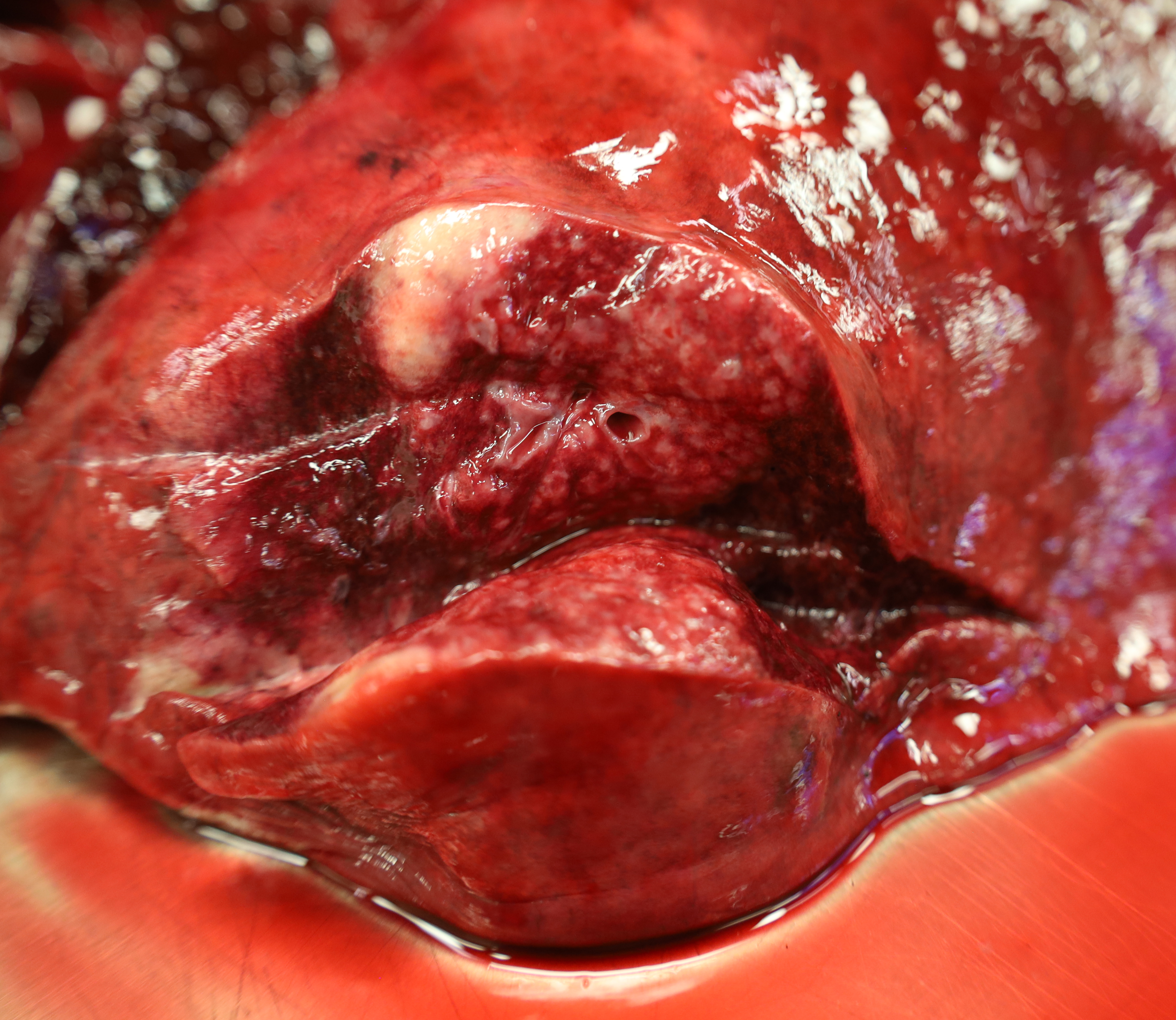
At necropsy examination, the distal trachea contained a small volume of stable tan foam. Approximately 20% of both left and right cranial lung lobes were firm, dark pink and well-demarcated from the surrounding parenchyma. On cut section, these areas exuded frothy white fluid and thin sections sank slightly in formalin before floating to the surface. Within the caudodorsal lung lobes there were ~5 indistinct multifocal firm nodules that ranged in size from 0.3 to 1.0 cm in diameter. On cut section these nodules were pale tan and merged with the surrounding parenchyma. Similar lung lesions were observed in other necropsied sheep from this cohort. One of the additional sheep also had unilateral suppurative mucoid yellow white fluid within the nasal passages and sinuses with diffuse reddening and slight thickening of the associated mucosa.
Laboratory Results :
Bacterial Culture Results: Lung grew 2+ Bibersteinia trehalose
PCR Results: Mycoplasma ovipneumoniae detected in lung tissue and by nasal swabs and confirmed through sequencing. Lung tissue was negative for bovine parainfluenza 3 virus, ovine progressive pleuropneumonia virus (Maedni Visna) and bovine respiratory syncytial virus.
Microscopic Description:
Two distinct patterns were observed within the examined sections of lung. Within caudodorsal regions (not apparent in submitted sections), there were large multifocal foci of mild histiocytic inflammation associated with nematodes of various life stages (adult, larval and eggs). Adult
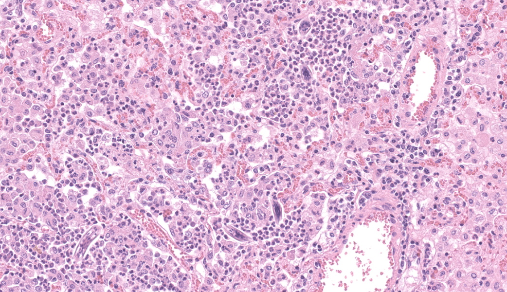
nematodes are ~80 mm in diameter and characterized by a thick outer eosinophilic cuticle, pseudocoelom, gastrointestinal tract lined by cuboidal epithelium with a brush border and reproductive tract that is often paired. Larvae are coiled and ~15 mm in diameter. Eggs are ~50mm in diameter and surrounded by a clear capsule containing eosinophilic granular material and variable numbers of nucleated cells. Nematodes fill the alveoli and are associated with small numbers of histiocytes, lymphocytes, and plasma cells. Lymphocytes and plasma cells sometimes form small aggregates or cuffs around blood vessels. Rare adult nematodes are observed within the larger airways. Minimal inflammation extends into the surrounding parenchyma.
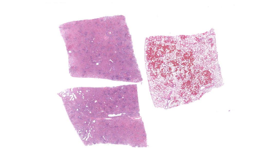
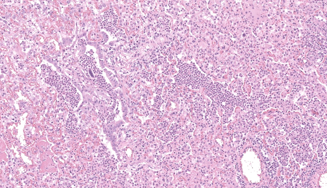
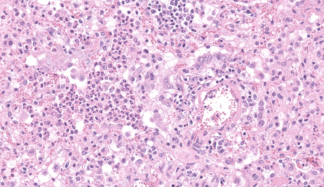
The second pattern (apparent in the submitted sections), observed within cranioventral sections of lung, is marked inflammation targeted towards small and large airways. Small terminal airways are most markedly affected and contain large numbers of luminal neutrophils. The surrounding respiratory epithelium is often hyperplastic and occasionally necrotic with sloughing into the airway lumen. Surrounding alveoli contain large numbers of luminal histiocytes with fewer neutrophils and occasionally are lined by cuboidal type II pneumocytes. There are rare multifocal areas of fibrin exudation into airways. Larval parasites resembling those described above are frequently seen individually within alveoli. Larger airways are often only minimally affected. Occasionally the lumen of smaller airways is compressed by nodular submucosal aggregates of inflammation (Figure 6). There are multifocal areas of acute hemorrhage throughout the lung (gunshot euthanasia associated).
Contributor's Morphologic Diagnosis:
Lung: Moderate, subacute, neutrophilic and histiocytic bronchopneumonia with epithelial hyperplasia
Lung, caudodorsal lobes: Mild, chronic multifocal histiocytic pneumonia with abundant intralesional nematodes and mild BALT hyperplasia
Contributor's Comment:
Histologic examination of the lungs confirmed a moderate bronchopneumonia as well as a mild lung worm infestation within the caudal lung lobes. A mild lymphoplasmacytic tracheitis was also present. Lung worms are ubiquitous in this bighorn sheep population and are most likely Protostrongylus stilesi, although P. rushii are also observed. Bacterial culture of the lung grew a mixed population including 2+ Bibersteinia trehalose, a bacterial respiratory pathogen capable of causing bronchopneumonia in bighorn sheep. PCR testing of the lung was positive for Mycoplasma ovipneumoniae (Movi). Due to the potential for cross reaction with other Mycoplasma spp. sequencing was also performed for confirmation. Lung tissue was also tested and was negative for common respiratory viruses including parainfluenza 3 virus, ovine progressive pleuropneumonia virus (Maedni Visna) and bovine respiratory syncytial virus. Due to the small size of this herd, the history of other animals coughing, and the potential for transmission to a larger nearby group of ewes and lambs, culling of the remaining rams was initiated. The majority of the additional rams that were necropsied exhibited bronchopneumonia similar to that already described.
Epizootic outbreaks of pneumonia have been a population limiting problem in bighorn sheep likely since the early 20th century and the expansion of humans and domestic small ruminants west across North America.3 Although pneumonia was recognized early as an important disease in wild bighorn sheep, and outbreaks were known to occur following exposures to domestic small ruminants, determining a definitive etiology took many years. Often, multiple potential respiratory pathogens were detected and therefore bighorn sheep pneumonia was discussed as a disease complex.1 Associated etiologies that were explored included lungworms (both P. rushii and P. stilesi), leukotoxin producing members of the Pasteurellaceae family (Mannheimia haemolytica, Bibersteinia trehalose and Pasteurella multocida) and Mycoplasma ovipneumoniae. Viral respiratory pathogens including parainfluenza virus 3 and respiratory syncytial virus were also occasionally detected, but less commonly.
Advanced molecular techniques and careful experimental infections eventually lead to the identification of Movi as the most significant and essential pathogen in epizootic pneumonia of bighorn sheep.2 Within domestic small ruminants, Movi is known as a cause of generally mild chronic bronchopneumonia in lambs, also known as atypical pneumonia or chronic non-progressive pneumonia.9 Movi can affect several non-domestic species of the Caprinae subfamily including bighorn sheep, Dall's sheep, mountain goats and muskox, and is often carried in the nasal passages of healthy domestic sheep and goats. 5,10 Outbreaks in bighorn sheep can often be traced to contact with domestic small ruminants, more often sheep than goats. Movi associated outbreaks in bighorn sheep occur in two distinct forms. The first introduction of a novel strain of Movi to a naive population leads to a slowly
progressing pneumonia that affects all age classes and often results in high morbidity and mortality rates of 30-90%.1 Reported clinical signs include coughing, nasal discharge, head shaking and ear drooping. The resultant pneumonia is frequently made more severe by secondary bacterial involvement often with members of the Pasteurellaceae such as the Bibersteinia trehalose observed in this case. Recurring outbreaks in subsequent years affect primarily lambs due to transmission from carrier ewes, and lead to low lamb recruitment and poor population growth. The outcome of infection in a population can be quite variable depending on complex interactions between host immunity, bacterial strain and environmental factors. Currently there are no vaccines or antibiotic therapies that have been found to be effective. 4
Movi pneumonia bears some resemblance to the atypical pneumonia of pigs caused by M. hyopneumoniae and shares several histologic and pathogenic features with other members of the Mycoplasma genus. Mycoplasma spp. target and destroy the cilia of airways impeding mucocilliary clearance and predisposing the lungs to secondary bacterial infection.1 Characteristic histologic features include hyperplasia of bronchiolar and alveolar epithelium, lymphoid (BALT) hyperplasia around airways and blood vessels, bronchiolar mucous metaplasia and more chronically, formation of distinct hyaline scars.7 Hyaline scars are a unique feature that bear some resemblance to bronchiolitis obliterans observed in chronic bovine pneumonias, including those with Mycoplasma bovis. Hyaline scars are described as nodular broad-based accumulations of brightly eosinophilic poorly cellular material below the respiratory epithelium that result in compression and distortion of the airway lumen.9 Unfortunately, depending on chronicity, many cases, including the current one, lack these characteristic histologic features and PCR testing and sequencing is necessary to confirm the diagnosis. In addition to bronchopneumonia, other Movi associated lesions include tracheitis, rhinitis, sinusitis and otitis media. 1
Management of these outbreaks requires an adaptive approach and can be quite challenging depending on the timing of detection and many other factors. 4 Preventative management should focus on minimizing contact between bighorn sheep and domestic small ruminants. In first incursion outbreaks, or when only a small herd is affected, complete depopulation may be an effective strategy given the short survival of the bacteria in the environment; Movi bacteria persist for only minutes outside of the host.4 In more chronically affected populations, management may focus on testing and removal of carrier individuals. In this particularly outbreak, culling of the small affected herd seems to have been effective in preventing spread to the surrounding populations.
Contributing Institution:
Diagnostic Services Unit (DSU) within the Faculty of Veterinary Medicine, University of Calgary. https://vet.ucalgary.ca/departments-units/dsu/home
JPC Morphologic Diagnosis:
Lung: Bronchopneumonia, suppurative and histiocytic, subacute, diffuse, marked, with mild BALT hyperplasia, early obliterating bronchiolitis, and numerous metastrongyle larvae.
JPC Comment:
This case was an excellent example of a real-world diagnostic case with multiple pathogens. This contributor provided a fantastic comment on Mycoplasma ovipneumonae (Movi) and Bibersteinia trehelosi, as well as gave an honorable mention to the ubiquitous lungworms Protostrongylus spp, in their write-up. Movi is the #1 infectious disease of Bighorn sheep.1 In cases of "bighorn sheep pneumonia", Movi is frequently the primary initiator of outbreaks and lays the foundation for a polymicrobial pneumonia.1
Bibersteinia trehelosi, previously Pasteurella trehelosi, is a common nasal commensal that is opportunistic in cases of bighorn sheep pneumonia.1,6 It is best known for its role in systemic pasteurellosis in 4-9mo lambs and is also considered one of the pathogens within the ovine respiratory complex.6
Participants discussed the potential identity of the nematodes and considered Dictyocaulus filaria, Muellerius capillaris, or Protostrongylus spp. Dictyocaulus spp were quickly ruled out as they are larger lungworms that usually reside in the upper airways.8 Muellerius capillaris was similarly discounted as they tend to accumulate in subpleural nodules.8 Protostrongylus spp, however, are small and tend to disperse diffusely throughout the pulmonary parenchyma, as seen in this case.8
The lungs here also highlighted nice examples of early obliterating bronchiolitis (also representing the continuing evolution of terminology, having been previously referred to as "bronchiolitis obliterans"), characterized by organizing aggregates of fibrin that are covered by bronchiolar epithelial cells within bronchiolar lumens.
References:
- Besser TE, Cassirer EF, Highland MA, Wolff P, Justice-Allen A, Mansfield K, Davis MA, Foreyt W. Bighorn sheep pneumonia: Sorting out the cause of a polymicrobial disease. Preventive Veterinary Medicine. 2013:108(2-3):85-93.
- Besser TE, Cassirer EF, Potter KA, Lahmers K, Oaks JL, Shanthalingam S, Srikumaran S, Foreyt WJ. Epizootic pneumonia of bighorn sheep following experimental exposure to Mycoplasma ovipneumoniae. PLOS ONE. 2014:9(10).
- Cassirer EF, et al. Pneumonia in bighorn sheep: Risk and resilience. Journal of Wildlife Management. 2018:82(1):32-45.
- Garwood TJ, Lehman CP, Walsh DP, Cassirer EF, Besser TE, Jenks JA. Removal of chronic Mycoplasma ovipneumoniae carrier ewes eliminates pneumonia in a bighorn sheep population. Ecology and Evolution. 2020:10(7):3491-3502.
- Handeland K, Tengs T, Kokotovic B, Vikoren T, Ayling RD, Bergsjo B, Sigurdardottir OG, Bretten T. Mycoplasma ovipneumoniae - a primary cause of severe pneumonia epizootics in the norwegian muskox (Ovibos moschatus) population. PLOS ONE. 2014:9(9).
- Jiang H, Yang L, Duan S, et al. Analysis of nasopharyngeal microbiota revealing microbial disturbance associated with ovine respiratory complex. Res Vet Sci. 2024;179:105383.
- Maxie MG. Jubb, Kennedy & Palmer's Pathology of Domestic Animals: Elsevier, 2016. 2:563-564.
- Miller DS, Weiser GC, Aune K, et al. Shared Bacterial and Viral Respiratory Agents in Bighorn Sheep (Ovis canadensis), Domestic Sheep (Ovis aries), and Goats (Capra hircus) in Montana. Vet Med Int. 2011;2011:162520.
- Sheehan M, Cassidy JP, Brady J, Ball H, Doherty ML, Quinn PJ, Nicholas RAJ, Markey BK. An aetiopathological study of chronic bronchopneumonia in lambs in Ireland. Veterinary Journal. 2007:173(3):630-637.
- Wolff PL, et al. Detection of Mycoplasma ovipneumoniae in pneumonic mountain goat (Oreamnos americanus) kids. Journal of Wildlife Diseases. 2019:55(1):206-212.