CASE IV:
Signalment:
Four-year-old Texel ewe, sheep (Ovis aries)
History:
In January 2018, a farm owner reported clinical signs of blindness and stumbling gait in three sheep. These sheep were part of a flock of 96 Texel sheep, ranging from 5 months to 7 years old. The sheep were raised in a semi-intensive grazing system. Sheep from different origins were purchased and introduced into the flock without any testing for infectious agents. The clinical course lasted approximately 2 months, and other clinical signs included anorexia and progressive weight loss. Due to poor prognosis, these three sheep were humanely euthanized and submitted for postmortem examination. One of the sheep is represented in this conference.
Gross Pathology:
At the gross examination, the ewe was in poor body condition. Sections of the cerebrum revealed multifocal to coalescing, irregular, yellow to brown, soft areas delimiting the periventricular region. The lungs were heavy and non-collapsed, with rib impressions, with a pale gray to pink color, and a diffusely rubbery texture. On the cut surface, multiple white and firm foci, measuring 0.1 to 0.5 cm in diameter were observed, mainly around bronchi. The mammary gland was mildly hardened. No significant lesions were observed in other organs, including multiple joints (stifle, carpi, and tarsi).
Laboratory Results:
The macerated brain and lungs of each animal were pooled, proviral DNA was isolated using a standard phenol-chloroform protocol (Sambrook and Russel, 2001), and the nested PCR was performed using a protocol that amplifies a 460 bp of 5'LTR region of proviral SRLV DNA (Ryan et al., 2000). The nested PCR for VMV was positive on the sheep's tissue pool (brain and lung).
Microscopic Description:
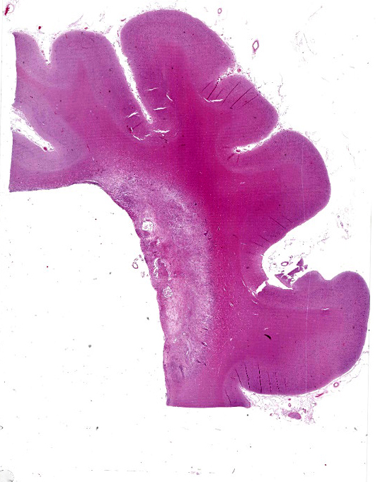
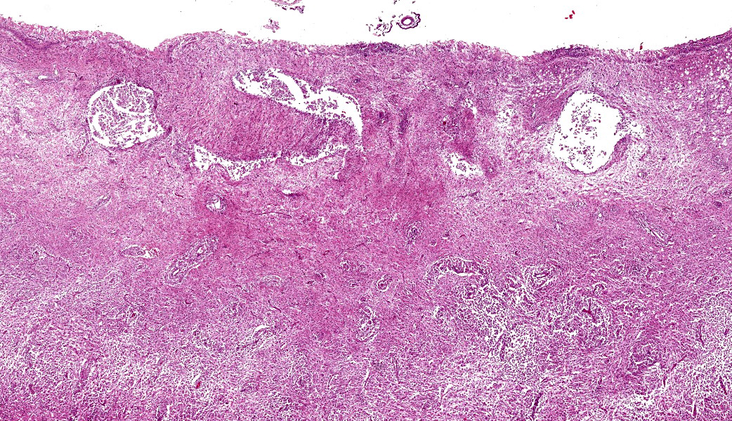
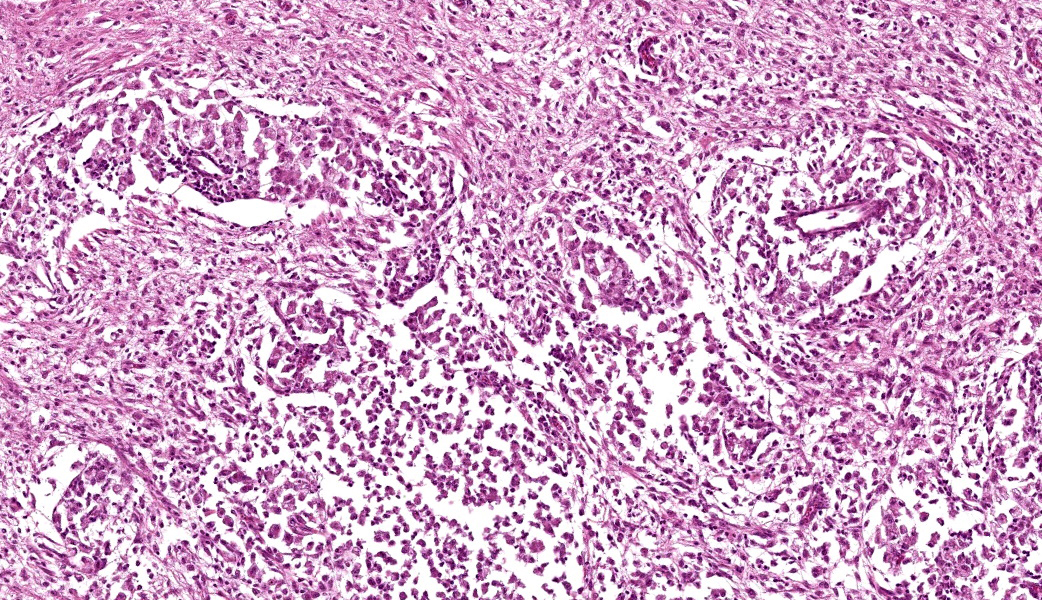
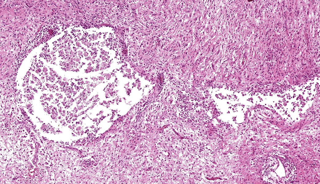
Cerebrum: In the white matter of the cerebrum, adjacent to the lateral ventricles, there is a well-demarcated, focally extensive area of marked neuropil rarefaction and liquefactive necrosis with numerous gitter cells and reactive astrocytes, including gemistocytes. Amidst this area, multifocal, marked inflammatory infiltrate of foamy macrophages and lymphocytes is observed, predominantly surrounding perivascular spaces, sometimes forming nodule-like structures. A similar inflammatory component is observed infiltrating, expanding, and partially effacing the adjacent ependymal epithelium. Additionally, cholesterol clefts, multifocal hemorrhage, multifocal white matter vacuolation, and mineral deposits are observed amidst this periventricular area of necrosis. The choroid plexus of the lateral ventricles (present in some slides) is expanded by abundant fibrin deposition, a small number of macrophages, and moderate to marked infiltrate composed mainly of lymphocytic aggregates forming lymphoid follicle-like structures. In the brainstem, the leptomeninges is moderately expanded by similar areas of perivascular infiltrate (not shown in the slides).
Contributor's Morphologic Diagnosis:
Cerebrum: Leukoencephalitis and ependymitis, necrotizing, lymphohistiocytic, focally extensive, marked, with demyelination, Texel, ovine.
Choroid plexus: Choroid plexitis, lymphohistiocytic, multifocal, marked, Texel, ovine.
Contributor's Comment:
Small ruminant lentiviruses (SRLV) are non-oncogenic retroviruses that infect sheep and goats, leading to diseases known as visna-maedi (VM; also known as ovine progressive pneumonia) and caprine arthritis and encephalitis (CAE).2 The etiological agents of VM and CAE are Visna-maedi virus (VMV) and caprine arthritis encephalitis virus (family Retroviridae, genus Lentivirus). The major tropism of SRLVs is for monocyte/macrophages and dendritic cells.2,9 Both diseases (VM and CAE) are multisystemic, progressive, and degenerative, with the establishment of persistent viral infection via viral genomic integration into the host's genome.1,9
Sheep infected with VMV develop a chronic wasting syndrome characterized by persistent infection in several organs, including lungs, central nervous system, mammary gland, and joints.1,10 The respiratory form (maedi) is characterized by lymphocytic interstitial pneumonia, smooth muscle hyperplasia and is the most common form of the disease.2 Clinical signs consist of progressive weight loss and dyspnea.1,2,10 Chronic progressive arthritis of the carpus or tarsus is seen in SRLV-infected animal, with a higher incidence in goats.2 A large proportion of sheep and goats infected by this virus present a non-suppurative, indurative mastitis.2 The neurologic form of the disease (visna) is sporadic and was first described in Iceland as non-suppurative demyelinating encephalitis clinically characterized by a chronic progressive paralytic disease of adult sheep.10 Different from other studies that describe that the lesions of the nervous form are mainly located in the white matter of the cerebellar peduncles, pons, and the medulla oblongata1. In our study the lesions were commonly observed extending from the periventricular areas, and choroid plexus. The occurrence of visna can be related to the involvement of neurotropic strains of VMV, and the severity of the lesions suggests that highly pathogenic strains of the virus are a probability.1
Clinical signs of visna are not pathognomonic; therefore, pathological and ancillary laboratory testing is necessary to differentiate this condition from those caused by other pathogens. Differential diagnoses for neurological disease in sheep include listeriosis, Border disease, rabies, louping-ill, polioencephalomalacia, coenurosis, and scrapie.4,7,8
The clinical features of rabies can be mistaken with the neurologic form of SRLV, as described in our cases, in which the sheep demonstrated similar neurologic signs. Nonetheless, the time lapse between the onset of clinical signs and death significantly differs between the two diseases. Rabies has acute clinical progression (approximately 5 days); visna is instead a chronic and progressive disease that can last for weeks or months.1,7
Contributing Institution:
Faculdade de Veterinaria Universidade Federal do Rio Grande do Sul Setor de Patologia Veterinaria http://www.ufrgs.br/patologia
JPC Morphologic Diagnosis:
Cerebrum: Periventricular demyelination, diffuse, severe, with liquefactive necrosis, hydrocephalus ex vacuo, and histiocytic leukoencephalitis and ependymitis.
JPC Comment:
This last case highlighted the classic neuropathologic presentation of the "visna" form of small ruminant lentivirus (SRLV) infection, and it prompted a rich discussion on lesion distribution, viral tropism, and breed susceptibility. "Maedi" is Icelandic for "dyspnea", referring to the respiratory form of SRLV infection, while "visna" means "shrinkage", describing the progressive neurologic wasting syndrome first recognized in Icelandic sheep. In the United States, the pulmonary form is more commonly known as ovine progressive pneumonia (OPP). Regardless of the clinical manifestation, the underlying agent is still SRLV, a non-oncogenic retrovirus with a major tropism for monocytes, macrophages, and dendritic cells.5,9
The histologic features in this case were striking. The dominant lesion was massive demyelination of the periventricular white matter, with relative sparing of the internal capsule. The presence of choroid plexus epithelium and lateral ventricle placed this section in the posterior telencephalon, consistent with the known predilection of small SRLV lesions for periventricular regions. Participants noted the flattened gyri and marked ventricular dilation, which are consistent with hydrocephalus ex vacuo. This is a form of hydrocephalus seen secondary to a loss of significant amounts of neuroparenchyma, which expands the ventricular system.
Dr. Brown emphasized that SRLV exemplifies a lifelong infection. Viral integration into the host genome allows the virus to persist silently for years before clinical disease emerges.5 Transmission patterns differ somewhat between syndromes: caprine arthritis encephalitis virus (CAE) is classically associated with ingestion of colostrum and milk transmission from infected ewes, whereas MVV is associated with spread via respiratory secretions among adult sheep.2 However, both viruses can use either route, and both establish chronic infection of macrophages.5 This macrophage tropism correlates well with the prominent histiocytic infiltrates in the CNS in this case.
The group briefly reviewed the clinical and epidemiologic implications of SRLV infection. Because infected animals remain lifelong carriers, seropositive sheep should be culled in control programs, and flock management must focus on preventing both respiratory spread and milk-borne transmission. The interplay between viral strain and host genetics determines whether an infected animal develops pulmonary "maedi", neurologic "visna", indurative mastitis, or chronic arthritis, and this Texel ewe exemplified the severe neurotropic form of the disease.
Texel sheep, as in this case, are among the breeds reported to be more susceptible to SRLV-associated disease. The reasons are not fully understood, but host genetics are thought to influence both viral replication and lesion severity.3 Texels have repeatedly been implicated in flocks with high disease burdens.3 Corriedales and Valais blacknose sheep are also reported to be more susceptible.3,6
References:
- Benavides J, Gomez N, Gelmetti D, Ferreras MC, Garcia-Pariente C, Fuertes M, Gircia-Marin JF, Perez V. Diagnosis of the nervous form of maedi-visna infection with a high frequency in sheep in Castilla y Leon, Spain. Vet Rec. 2006;158:230-235.
- Blacklaws B. Small Ruminant Lentiviruses: Immunopathogenesis of visna-maedi and caprine arthritis and encephalitis virus. Comp Immunol Microbiol Infect Dis. 2012;35:259-269.
- de Miguel R, Arrieta M, Rodriguez-Largo A, Echeverria I, Resendiz R, Perez E, Ruiz H, Perez M, de Andres D, Reina R, de Blas I, Lujan L. Worldwide Prevalence of Small Ruminant Lentiviruses in Sheep: A Systematic Review and Meta-Analysis. Animals (Basel). 2021 Mar 11;11(3):784.
- Guedes KMR, Riet-Correa F, Dantas AFM, Simoes SVD, Miranda Neto EG, Nobre VMT, Medeiros RMT. Diseases of the central nervous system in goats and sheep of the semiarid. Pesq Vet Bras. 2007;27(1):29-38.
- Kalogianni AI, Stavropoulos I, Chaintoutis SC, Bossis I, Gelasakis AI. Serological, Molecular and Culture-Based Diagnosis of Lentiviral Infections in Small Ruminants. Viruses. 2021 Aug 27;13(9):1711.
- Letko A, Butzberger C, Hirter N, Paris JM, Abril C, Drogemuller C. Genetic evaluation of small ruminant lentivirus susceptibility in Valais blacknose sheep. Anim Genet. 2021 Oct;52(5):781-782.
- Rissi DR, Pierezan F, Kommers GD, Barros CSL. Occurrence of rabies in sheep in Rio Grande do Sul, Brazil. Pesq Vet Bras. 2008;28(10):495-500.
- Rissi DR, Pierezan F, Oliveira Filho JC, Fighera RA, Irigoyen LF, Kommers GD, Barros CSL. Diseases of sheep from central Rio Grande do Sul State, Brazil: 361 cases. Pesq Vet Bras. 2010;30(1):21-28.
- Ryan S, Tiley L, McConnell I, Blacklaws B. Infection of dendritic cells by the Maedi-Visna Lentivirus. J Virol. 2000;74(21):10096-10103.
- Sigurdsson B, Palsson PA, Grimsson H. Visna, a demyelinating transmissible disease of sheep. J Neuropathol Exp Neurol. 1957;16:389-403.