CASE I:
Signalment:
22 month-old, neutered male, dog, Schnauzer.
History:
The dog was referred to the Veterinary Hospital with a history of weight loss, inappetence, vomiting, and mild diarrhea. Blood samples were collected for total blood cell counts, a biochemical profile, and serological tests to detect anti-Leishmania antibodies. Blood samples were collected at admission and weekly while the dog was under medical care. Physical examination demonstrated enlargement of the cervical and popliteal lymph nodes. Clinical examination also revealed abdominal pain, hepatomegaly, and splenomegaly. Ultrasonography confirmed splenomegaly and demonstrated intestinal wall thickening. Aspirative cytology evaluation of the spleen and popliteal lymph node was performed. The first blood cell count revealed anemia, thrombocytopenia, and mild monocytosis. Based on the hematology results, erlichiosis was suspected. Based on initial suspicion, doxycycline (5 mg/kg IV bid), maropitant citrate (1 mg/kg sc for 5 d), and fluid (ringer lactate) were prescribed. Therefore, an exploratory laparotomy with splenectomy was performed. The spleen was submitted to histopathology. A re-check five days after the onset of therapy showed no changes on clinical examination. Cytopathological findings were compatible with histiocytic lymphadenitis and splenitis associated with higher numbers of cell-associated acid-fast bacilli. Then, 20 days after admission, the dog was treated with metronidazol (15 mg/kg, IV, bid, 7 days), amoxicillin and clavulanate (20 mg/kg, IV, bid, 14 days), silymarin (25 mg/kg, vo, sid, 14 dias), ursodeoxycholic acid (15 mg/kg, vo, sid 10 days), ondansetron (0.22 mg/kg, IV, tid, 14 days), and rifampicin (20 mg/kg, vo, tid) used to treat Mycobacterium infection. Ten days later, the clinical condition worsened, and the dog presented with regurgitation of food, weakness, and lethargy. The dog was monitored since admission, totaling 30 days, when he died after rapid deterioration of his clinical condition. The owner authorized a cosmetic necropsy. Most organs were collected for histopathology. Samples of spleen and lymph nodes were collected and frozen for molecular analysis.
Gross Pathology:
The spleen (surgically removed) was markedly enlarged, with roughly twice its normal size. The subcapsular surface presented numerous multifocal to coalescing yellow-whitish foci. Approximately 90% of the parenchyma was replaced by multifocal to coalescing prominent, slightly soft, yellow-whitish millimetric areas. Two focally extensive whitish, amorphous, and soft areas measuring approximately 4 cm in diameter were grossly interpreted as caseous necrosis. At necropsy, the dog was emaciated, and the oral and ocular mucosae were white to slightly yellow. All cervical, axillary, popliteal, mediastinal, tracheobronchial, iliac (external and internal), and mesenteric lymph nodes were markedly enlarged, some of which were more than five times their normal size. The capsules of these lymph nodes were tense, and on the cut surface their normal morphologic characteristic were lost, with the cortex and medulla replaced by yellow-whitish solid soft tissue. The ventral portions of the pulmonary lobes (especially on the left side) were dark-red and consolidated, with viscous yellow exudate draining from the bronchial lumen and parenchyma. The liver was markedly enlarged, with rounded edges and a yellowish surface. Filaments and plaques of fibrin were observed on the capsular surface of the right hepatic lobes. On cut surface, multifocal to coalescing slightly prominent yellow-whitish foci were interspersed with red-yellowish parenchyma. Kidneys were pale red, interspersed with several whitish soft foci in the cortex.
Laboratory Results:
Serological tests for Leishmania: Indirect immunofluorescent (RIFI) in 1:40 dilution and enzyme-linked immunosorbent assay (ELISA) for IgG were negative.
Clinical pathology:
CBC: 4.79 Hgb, 9.7 Hgb; 28.0 % Hct; 12.912 Seg; 1.603 Mno; 166.000 platelets and metarubricytes, 2.9%.
Biochemical profile: alkaline phosphatase, 552.0 U/L; ALT, 177.35 U/L; AST, 223.73 U/L. Albumin, 1.69 g/dL Popliteal lymph node aspirate biopsy: many macrophages with numerous small vacuoles in the cytoplasm.
Acid-fast (Ziehl-Neelsen) staining of lymph node and spleen aspirates: large numbers of acid-fast bacilli associated with macrophages and extracellular
Molecular analysis of DNA extracted from spleen and feces:
PCR for M. bovis, M. tuberculosis and M. avium subsp. resulted negative.
PCR and sequencing identified the bacilli as Mycobacterium avium subsp. avium.
Histopathologic Description:
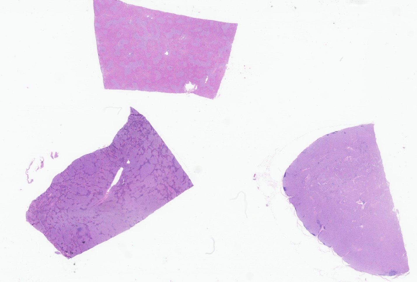
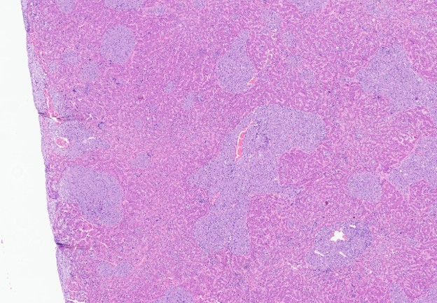
Histopathological evaluation of the spleen revealed marked inflammatory reaction replacing the normal architecture. Multifocal to coalescing aggregates of epithelioid macrophages and fewer multinucleated cells compressed and replaced the red and white pulp. The remaining normal red pulp had several nucleated erythroid cells and some megakaryocytes. Lymphocytes and plasma cells were markedly reduced. In addition, a focally extensive area consisted of amorphous eosinophilic material mixed with fibrin and cellular debris.
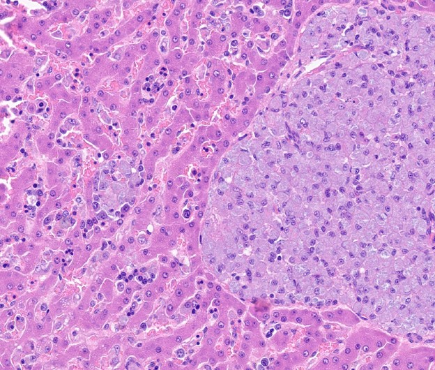
The lymph node parenchyma was 90 to 95% replaced by macrophages, epithelioid macrophages, and a lower number of multinucleate cells (2-3 nuclei). The remaining parts of the architecture were the subcapsular sinus and the small lymphoid follicles. Inflammatory cells similar to those in the spleen and lymph nodes replaced about 50-60% of the normal liver parenchyma. In the sinusoids of these are as, numerous lymphocytes, several nucleated erythroid cells, and fewer megakaryocytes were present.
The mucosa and submucosa of the small and large intestine were thickened by granulomatous inflammation, composed mostly of epithelioid macrophages and a lower number of multinucleated cells with up to five nuclei. In the small intestine, granulomatous inflammation replaced 90% of the lymphoid follicles (Peyer patches).
Pulmonary lobes showed suppurative bronchopneumonia and fibrinosuppurative pleuritis. In addition, there was an occlusive thrombus in a bronchial artery. Multifocal interstitial and alveolar granulomatous inflammation was also present.
The kidneys had mild multifocal interstitial granulomatous inflammation, similar to that observed in the other organs mentioned above. In addition, moderate membranous glomerulopathy and tubular proteinuria were present.
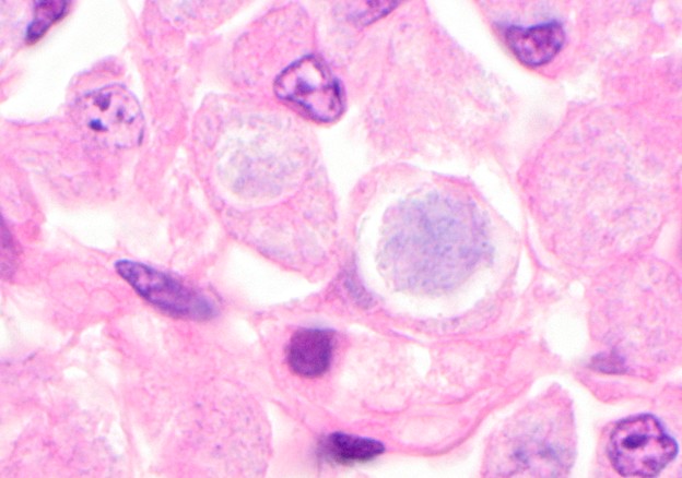
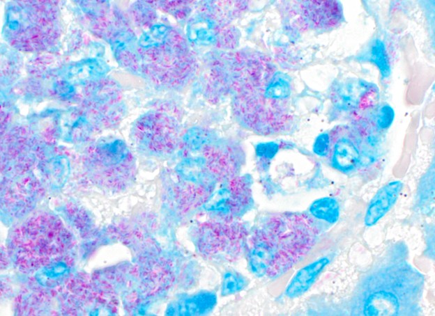
Ziehl-Neelsen special staining of the spleen, lymph nodes, liver, lungs, kidneys, and intestine showed large amounts of acid-fast bacilli within macrophages and multinucleated cells.
Contributor's Morphologic Diagnoses:
Spleen: marked multifocal to coalescing granulomatous splenitis, lymphoid atrophy, locally extensive lytic necrosis, and moderate extramedullary hematopoiesis.
Lymph nodes: marked diffuse granulomatous lymphadenitis and lymphoid atrophy.
LIver: marked multifocal to coalescing granulomatous hepatitis and moderate extramedullary hematopoiesis.
Not submitted tissues:
Small and large intestine (mucosa and submucosa): moderate multifocal to coalescing circumferential granulomatous enteritis and lymphoid atrophy in the gut-associated lymphoid tissue.
Lungs: marked acute suppurative broncopneumonia, and mild multifocal granulomatous pneumonia.
Kidneys: mild multifocal granulomatous nephritis and moderate membranous glomerulopathy associated with tubular proteinuria.
Contributor's Comment:
Gross and histopathological findings associated with acid-fast positive stain were compatible with disseminated mycobacteriosis. The large numbers of intracellular bacilli are more suggestive of mycobacteriosis caused by M. avium. Molecular analysis, including sequencing, confirmed M. avium subsp. avium infection in this dog. The three most common species of Mycobacterium, namely M. tuberculosis (human), M. bovis (cattle), and M. avium (avian species), affect their preferential hosts more often. Still, cross-infections may occur involving several other host species. M. bovis and M. tuberculosis cause tuberculosis characterized by disseminated or localized infections in the lungs or in the gastrointestinal tract. M. avium, one of the species of the M. avium-intracellulare complex (MAC), can cause mycobacteriosis or atypical mycobacterium4. The avium species includes 4 subspecies, i.e., M. avium subsp. avium, M. avium subsp. hominissuis, M. avium subsp. silvaticum, and M. avium subsp. paratuberculosis2, 16. M. avium subsp. avium infection is common in domestic11, wild8, and exotic captive birds 5, but it is rare in mammals1. M. genavense is another common cause of mycobateriosis in birds12. In dogs, disseminated mycobacteriosis has also been caused by M. avium subspecies avium7,1 and M. avium subspecies hominis9,3. Disseminated canine tuberculosis caused by M. tuberculosis infection has also been reported13.
Lesions produced by M. avium infection are more proliferative and consist of diffuse infiltration of macrophages, epithelioid cells, and multinucleate cells, accompanied by numerous bacilli and extensive fibroplasia. Dogs and cats usually present lesions as granulation tissue4. The disseminated infection in this dog was characterized by diffuse granulomatous inflammation (predominantly macrophages and epithelioid cells, with occasional giant cells) with numerous bacilli, similar to M. avium infection in birds. Caseous necrosis was present only in two locally extensive areas in the spleen. Similar findings caused by M. avium were described in dogs7 and in cats10. A genetic predisposition to MAC infections may exist in basset hounds, Siamese cats10 and in schnauzers7. Eggers et al.7detected depletion of T and B lymphocytes in three miniature schnauzers with disseminated mycobacteriosis. Evidence suggesting or corroborating immunosuppression could not be confirmed in this dog. Nevertheless, another fact should be considered. This dog was the only infected animal in a house with three other animals. The dog lived permanently in the backyard with three other animals: a dog and two cats. One cat died almost at the same time as this dog; however, it was diagnosed with pulmonary carcinoma. Frozen tissue of this cat was tested for Mycobacterium spp. and resulted negative. The dog and another cat were monitored during four months, and no abnormalities were detected. Feces of these animals were collected twice and tested by culture and PCR for Mycobacterium spp. and were negative.
The cause of progressive anemia was not well defined. Renal lesions were considered insufficient to interfere with erythropoietin and produce anemia. Intestinal lesions could impair nutrient absorption, and a possible effect on red blood cell production may occur. Nevertheless, dissemination of macrophages loaded with myriads of bacterial rods throughout bone marrow could determine myelophthisis. The bone marrow was not examined in this case, but its involvement and consequent anemia in schnauzers with disseminated mycobacteriosis have been described14,15.
The gastrointestinal route of infection could be suspected because the lesions were predominant in the gastrointestinal organs and less intense in the respiratory tract. The source of infection was possibly drinking water, since the owner reported that, some weeks before the animal presented with signs of disease, he found wild bird feces in the dog's drinking water. The backyard had some trees and birds (especially sparrows). Wild birds (sparrows, crows, and pigeons) may act as reservoirs and can shed the infection to the environment17. The bacteria can persist in the environment for many years, especially in the soil5,6.
MAC is an opportunistic pathogen in immunocompromised humans2. The dog was in close contact with its owners, but they had no history of disease.
Contributing Institution:
Veterinary School
Universidade Federal de Minas Gerais
www.vet.ufmg.br
JPC Diagnoses:
- Liver: Hepatitis, granulomatous, chronic, multifocal to coalescing, severe, with numerous intracytoplasmic bacilli.
- Spleen: Splenitis, granulomatous, chronic, diffuse, severe, with numerous intracytoplasmic bacilli.
- Lymph node: Lymphadenitis, granulomatous, chronic, diffuse, severe, with numerous intracytoplasmic bacilli.
- Liver, spleen: Extramedullary hematopoiesis, diffuse, mild to moderate.
JPC Comment:
All clinical presentations of mycobacteriosis are uncommon in dogs, and disseminated disease is even rarer4. An interesting feature of mycobacteriosis in both dogs and cats is an apparent breed predisposition. In dogs, miniature schnauzers, as in this case, and basset hounds are predisposed. In cats, reported predispositions include the Siamese, Somali, and Abyssinian breeds4,20. These breed associations suggest underlying genetic factors, likely involving genes that impair the host's ability to recognize and neutralize Mycobacterium species.
The miniature schnauzer has the best-characterized genetic defect. Miniature schnauzers at increased risk of mycobacteriosis frequently harbor a CARD9 deficiency caused by a mutation in the CARD9 gene4, 19, 21. CARD9 is an adapter signaling protein best known for its critical role in antifungal immunity. Interestingly, humans with analogous CARD9 deficiencies do not appear predisposed to mycobacterial infections; however, as expected, they face a heightened risk of severe fungal disease21. Notably, these patients often develop localized or systemic Candida infections without other predisposing conditions, a presentation characteristic of CARD9 deficiency19. Mechanistically, fungi are recognized by various pattern recognition receptors (PRRs), which recruit CARD9 to trigger the host innate immune response21. Although the presentation differs between humans and dogs, absent or minimal activation of the host's innate immune system against organisms that are generally nonpathogenic in healthy, immunocompetent individuals is a key unifying feature.
References:
- Bauer N, Burkhardt S, Kirsch A, Weiss R, Moritz A, Baumgaertner W. Lymphadenopathy and diarrhea in a Miniature Schnauzer. Vet Clin Pathol. 2002;31:61-64.
- Biet F, Boschiroli ML, Thorel MF, Guilloteau LA. Zoonotic aspects of Mycobacterium bovis and Mycobacterium avium-intracellulare complex (MAC). Vet Res. 2005;36:411-436.
- Campora L, Corazza M, Zullino C, Ebani VV, Abramo F. Mycobacterium avium subspecies hominissuis disseminated infection in a Basset Hound dog. J Vet Diagn Invest. 2011;23:1083-1087.
- Caswell JL, Willians KJ. Respiratory system. In: Jubb, Kennedy & Palmer's Pathology of Domestic Animals, ed. Maxie MG, 5th ed., Toronto, Canada: Elsevier Saunders; 2007:524-650.
- Del Pilar Silva A, Leon CI, Guerrero MI, Neira R, Arias L, Rodriguez G. Avian tuberculosis of zoonotic importance at a zoo on the Bogota Andean plateau (Sabana), Colombia. Can Vet J. 2009;50:841-845.
- Dhama K, Mahendran M, Tiwari R, Dayal S, Kumar D, Sawant PM.
- Tuberculosis in Birds: Insights into the Mycobacterium avium Vet Med Int. 2011. 2011: 712369.
- Eggers JS, Parker GA, Braaf HA, Mense MG. Disseminated Mycobacterium avium infection in three Miniature Schnauzer littermates. J Vet Diagn Invest. 1997;9:424-427.
- Quesada-Canales O, Diaz-Delgado J, Paz Y, Dominguez L, Bezos J, Calabuig P, et al. Disseminated avian mycobacteriosis in a free-living grey heron (Ardea cinerea). Avian Dis. 2013;57:703-706.
- Haist V, Seehusen F, Moser I, Hotzel H, Deschl U, Baumgartner W, et al. Mycobacterium avium hominissuis infection in 2 pet dogs, Germany. Emerg Infect Dis. 2008;14:988-990.
- Jordan HL, Cohn LA, Armstrong PJ. Disseminated Mycobacterium avium complex infection in three Siamese cats. J Am Vet Med Assoc. 1994;204:90-93,
- Kriz P, Kaevska M, Bartejsova I, Pavlik I. Mycobacterium avium subsp. avium found in raptors exposed to infected domestic fowl. Avian Dis. 2013;57:688-692.
- Manarolla G, Liandris E, Pisoni G, Sassera D, Grilli G, Gallazzi D, et al. Avian mycobacteriosis in companion birds: 20-year survey. Vet Microbiol. 2009;133:323-327.
- Martinho APV, Franco MMJ, Ribeiro MG, Perrotti IBM, Mangia SH, et al. Case Report: Disseminated Mycobacterium tuberculosis infection in a dog. Am J Trop Med Hyg. 2013; 88:596-600.
- Miller MA, Greene CE, Brix AE. Disseminated Mycobacterium avium-intracellulare complex infection in a miniature schnauzer. J Am Anim Hosp Assoc. 1995;31:213-216.
- O'Toole D, Tharp S, Thomsen BV, Tan E, Payeur JB. Fatal mycobacteriosis with hepatosplenomegaly in a young dog due to Mycobacterium avium. J Vet Diagn Invest. 2005;17:200-204.
- Rindi L, Garzelli C. Genetic diversity and phylogeny of Mycobacterium avium. Infect Genet Evol. 2014;21:375-383.
- Tell LA, Woods L, Cromie RL. Avian tuberculosis in birds, Review Science and Technology. Office Internationale des Epizooties. 2001;20: 180-203.
- Ghielmetti G, Giger U. Mycobacterium avium: an Emerging Pathogen for Dog Breeds with Hereditary Immunodeficiencies. Curr Clin Microbiol Rep. 2020;7(3):67-80.
- Sykes JE. Cutaneous Mycobacterioses of Cats and Dogs. Vet Clin North Am Small Anim Pract. 2025;55(2):237-249.
- Drummond RA, Franco LM, Lionakis MS. Human CARD9: A Critical Molecule of Fungal Immune Surveillance. Front Immunol. 2018;9:1836.