Joint Pathology Center
Veterinary Pathology Services
Wednesday Slide Conference
2017-2018
Conference 25
May 9th, 2018
CASE IV: Case 2 (JPC 4101147)
Signalment: 3.5-month-old, female, CyBB knockout (Mus musculus), mouse.
History: The animal had been used as a breeder and was found dead. Dystocia was the top differential diagnosis.
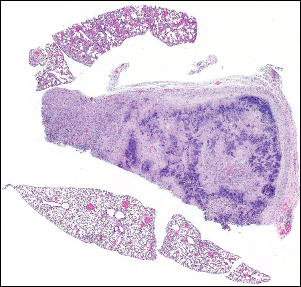
Gross Pathology: There is a mass replacing the caudal right lung lobe that contains inspissated white-tan material (abscess), with few scattered tan pinpoint-1mm foci throughout all lobes. There is bloody discharge from the vulva. The uterus is gravid with 2-3 pups in each horn. The spleen is enlarged 5- fold.
Laboratory results: Bacterial culture yielded positive growth for Klebsiella pneumoniae ssp pneumonia from heart blood, as well as swabs from the lung abscess.
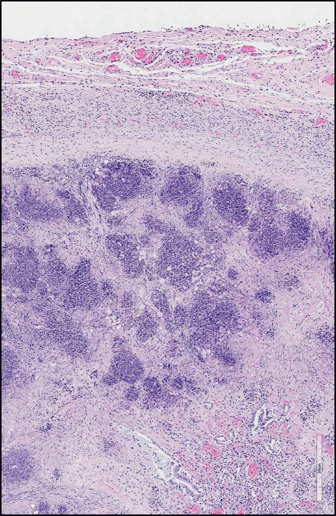
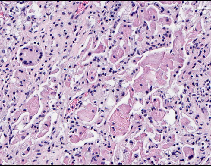
Microscopic Description: Lung: There is marked expansion and replacement of architecture by a central region of lytic cell debris, as well as lytic and degenerate neutrophils surrounded by an outer layer of collagen and fibroblasts (abscess). Within some sections, where inflammation is less extensive, there are alveoli lined by cuboidal epithelium (type 2 pneumocyte hyperplasia), and are filled with degenerate neutrophils, epithelioid macrophages and multinucleated giant cells with variably-sized eosinophilic rhomboid crystals. There is compression of adjacent relatively normal parenchyma, where alveoli are filled with similar inflammatory cells containing crystals, which are also present in large numbers in peripheral alveolar spaces in non-abscessed lung sections. Similar crystals are also present free within the lumens of airways. The associated pleura is expanded by immature fibrous connective tissue, macrophages, lymphocytes and plasma cells, and congested small blood vessels.
Contributor’s Morphologic Diagnosis:
Lung: Multifocal to coalescing suppurative pneumonia with abscesses; Multifocal to coalescing granulomatous pneumonia with intracytoplasmic eosinophilic crystals (acidophil macrophage pneumonia)
Contributor’s Comment: The submitted slide illustrates two relatively classic lesions seen in mice: bacterial pneumonia with abscessation and acidophil macrophage pneumonia. Klebsiella pneumoniae is an enteric commensal bacterium and is considered an opportunistic pathogen. It has been associated with suppurative lesions of the female reproductive tract in aged B6C3F mice, as well as perianal dermatitis, preputial abscesses, otitis and bacteremic disease (leading to pneumonia and abscesses in other organs) in mice of all ages and strains.1 Although not shown, this animal also had necrosuppurative metritis and similar inflammation affecting numerous organs, indicating bacteremia.
Acidophil macrophage pneumonia (AMP), also known as eosinophilic crystalline pneumonia, typically occurs in older animals with B6, 129 and Swiss mice being more susceptible.1 These crystals have been shown to be composed of iron, α-1 antitrypsin, immunoglobulin and granulocytic breakdown products, as well as Ym1 chitinase.1,5 AMP is typically an incidental finding, but can cause mortality in severe cases.1,3
Both bacterial pneumonia and acidophil macrophage pneumonia, along with splenomegaly, are documented to occur in the present genetically engineered mouse (homozygous null CyBB, or cytochrome b-245, beta polypeptide).2 This transgenic mouse is used to model chronic granulomatous disease in humans and is considered to be highly susceptible to bacterial infections.2,4 These mice are also highly susceptible to fungal infections; however, none were observed on H&E stained slides.
JPC Diagnosis: 1. Lung: Pneumonia, necrosuppurative, multifocal to coalescing, severe.
- Lung, Pneumonia, granulomatous, diffuse, moderat, with numerous intrahistiocytic intracytoplasmic, eosinophilic crystals.
Conference Comment: Klebsiella spp. are common commensal bacteria of the gastrointestinal tract of mice in the family Enterobacteriae. These bacteria, although usually quiescent, have the ability to become opportunistic pathogens. In mice (particularly immunodeficient mice), several disease processes have been linked to Klebisella spp. including: suppurative endometritis, perianal dermatitis, preputial abscesses, otitis, tooth infections, urogenital infections, pneumonia, and bacteremic disease (cervical lymphadenopathy, hepatic and renal abscesses, empyema, myocarditis, and thrombosis). Lesions are not characteristic and diagnosis is dependent upon virus isolation with either Klebsiella pneumoniae or K. oxytoca being the most common isolates.1
Acidophilic macrophage pneumonia (AMP) is characterized by accumulation of eosinophilic crystals within macrophages and free within alveolar spaces and is often an incidental finding in certain strains of mice (listed by the contributor above). In severely affected mice, the lungs are grossly red to tan and do no collapse. Ultrastructurally, the crystals are needle-shaped or rhomboidal which a clear lattice structure which repeats every 3-5 nm and are quite complex, composed of Ym1 and Ym2 chitinase, containing iron, alpha-1 antitrypsin, immunoglobulin, and breakdown products of granulocytes. This ultrastructural appearance is similar to Charcot-Leyden crystals associated with eosinophil-rich diseases in humans and non-human primates. Older mice frequently have a small number of alveolar macrophages containing crystals which become pathologic when a disease process impairs clearance of alveolar exudate and the crystal-laden macrophages accumulate and fill vital air spaces leading to dyspnea. Other locations in predisposed mice also contain Ym1 and Ym2 chitinase including: epithelium of the olfactory, nasal respiratory, middle ear, trachea, lung, stomach, gall bladder, bile duct, and pancreatic duct.1
Conference participants were hesitant to describe this lesion as an abscess because it is lacking a definitive capsule and there is normal tissue in between areas of lytic necrosis, preferring this to represent multifocal to coalescing areas of lytic necrosis.
Contributing Institution:
http://www.yerkes.emory.edu/research/divisions/pathology/index.html
References:
- Barthold SW, Griffey SM, Percy DH. Pathology of Laboratory Rodents and Rabbits. 4th Ames, IA: Blackwell Publishing Professional; 2007:62, 94-95.
- Bingel SA. Pathology of a mouse model of x-linked chronic granulomatous disease. Contemp Top Lab Anim Sci. 2002; 41(5):33-38.
- Hoenerhoff MJ, Starost MF, Ward JM. Eosinophilic crystalline pneumonia as a major cause of death in 129S4/SvJae mice. Vet Pathol. 2006; 43(5):682-688.
- Pollock JD, Williams DA, Gifford MA, Li LL, Du X, Fisherman J, Orkin SH, Doerschuk CM, Dinauer MC. Mouse model of X-linked chronic granulomatous disease, an inherited defect in phagocyte superoxide production. Nat Genet. 1995; 9(2):202-209.
- Ward JM, Yoon M, Anver MR, Haines DC, Kudo G, Gonzalez FJ, Kimura S. Hyalinosis and Ym1/Ym2 gene expression in the stomach and respiratory tract of 129S4/SvJae and wild-type and CYP1A2-null B6, 129 mice. Am J Pathol. 2001; 158(1):323-332.