Signalment:
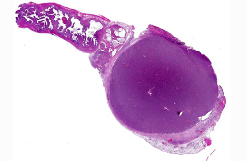
Gross Description:
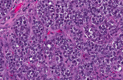
Histopathologic Description:
Morphologic Diagnosis:
Condition:
Contributor Comment:
In humans, granulosa cell tumors represent 2-5% of all ovarian tumors and usually have clinical features of hyperestrogenism.(16) Mutations in transcription factor FOXL2 is pathognomonic for adult-type granulosa cell tumors in humans.(17) Further, 17β-estradiol, inhibin, and mullerian inhibiting substance are considered to be reliable diagnostic and prognostic markers in humans.(8) While inhibin-α is a reliable marker in canine granulosa cell tumors(14) calretinin, GATA-4, and neuron-specific enolase are consistently expressed in non-human primate granulosa cell tumors.(3) No such markers have been reported in Syrian hamsters.
JPC Diagnosis:
1. Ovary: Granulosa cell tumor.
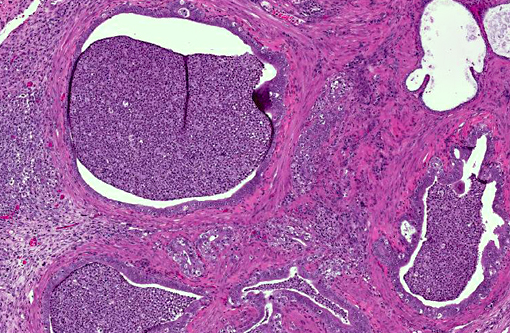
2. Oviduct: Salpingitis, suppurative, diffuse, moderate, with ductular ectasia.
Conference Comment:
The interesting aspect of sex cord-stromal tumors is their ability to produce a variety of hormones and induce corresponding clinical signs. The contributor mentioned those exhibited by mares, and recent publications have demonstrated increased levels of testosterone and inhibin can be useful in obtaining a presumptive diagnosis.(4) Additionally, anti-M+â-+llerian hormone concentrations are elevated in mares and cows with these neoplasms.(1,4) Estrogen and progesterone may also be secreted, as cystic endometrial hyperplasia and pyometra are common in bitches with sex cord-stromal tumors.(15) In this case, we believe the oviduct and uterus are moderately distended, and there is suppurative inflammation in some sections within the oviduct. The relationship of these inflammatory changes with the ovarian neoplasm is unclear.
References:
1. Ball BA, Almeida J, Conley AJ. Determination of serum anti-Mullerian hormone concentrations for the diagnosis of granulose-cell tumours in mares. Equine Vet J. 2013;45(2):199-203.
2. Beamer WG, Hoppe PC, Whitten WK: Spontaneous Malignant Granulosa Cell Tumors in Ovaries of Young SWR Mice. Cancer Res. 1985:45(11 Part 2):5575-5581.
3. Durkes A, Garner M, Juan-Sall+â-¬s C, Ramos-Vara J: Immunohistochemical Characterization of Nonhuman Primate Ovarian Sex CordStromal Tumors. Vet Pathol. 2012:49(5):834-838.
4. Gee EK, Dicken M, Archer RM, Herdan CL, Pauwels FE, Drayton BM. Granulosa theca cell tumour in a pregnant mare: concentrations of inhibin and testosterone before and after surgery. N Z Vet J. 2012;60(2):160-163.
5. Gelberg HB, McEntee K: Feline Ovarian Neoplasms. Vet Pathol. 1985:22(6):572-576.
6. Gregson RL, Lewis DJ, Abbott DP: Spontaneous ovarian neoplasms of the laboratory rat. Vet Pathol. 1984:21(3):292-299.
7. Kennedy PC, Cullen JM, Edwards JF, et. al. Histological Classification of Tumors of the Genital System of Domestic Animals. 2nd series, Vol. 4. Washington, D.C.: Armed Forces Institute of Pathology; 1998:24-25.
8. Kondo H, Onuma M, Shibuya H, Sato T: Spontaneous tumors in domestic hamsters. Vet Pathol. 2008:45(5):674-680.
9. Kottarathil VD, Antony MA, Nair IR, Pavithran K: Recent advances in granulosa cell tumor ovary: a review. Indian J Surg Oncol. 2013:4(1):37-47.
10. Maclachlan NJ, Kennedy PC: Tumors of the Genital Systems, Sex-cord stromal tumors. In: Meuten DJ, ed. Tumors in Domestic Animals. 4 ed. Ames, IA: Iowa State Press; 2002: 550-551.
11. McInnes EF, Ernst H, Germann P-G: Spontaneous Neoplastic Lesions in Control Syrian Hamsters in 6-, 12-, and 24-Month Short-Term and Carcinogenicity Studies. Toxicol Pathol. 2012.
12. Patnaik AK, Greenlee PG: Canine Ovarian Neoplasms: A Clinicopathologic Study of 71 Cases, Including Histology of 12 Granulosa Cell Tumors. Vet Pathol.1987:24(6):509-514.
13. Pour P, Mohr U, Althoff J, Cardesa A, Kmoch N: Spontaneous tumors and common diseases in two colonies of Syrian hamsters. III. Urogenital system and endocrine glands. J Natl Cancer Inst. 1976:56(5):949-961.
14. Riccardi E, Greco V, Verganti S, Finazzi M: Immunohistochemical Diagnosis of Canine Ovarian Epithelial and Granulosa Cell Tumors. J Vet Diagn Invest. 2007:19(4):431-435.
15. Schlafer DH, Miller RB: Female genital system. In: Maxie MG, ed. Jubb, Kennedy, and Palmer's Pathology of Domestic Animals. St. Louis, MO: Saunders Elsevier; 2007: 450-451.
16. Schumer ST, Cannistra SA: Granulosa Cell Tumor of the Ovary. J Clin Oncol. 2003:21(6):1180-1189.
17. Shah SP, K+â-¦bel M, Senz J, Morin RD, Clarke BA, Wiegand KC, et al.: Mutation of FOXL2 in Granulosa-Cell Tumors of the Ovary. N Engl J Med. 2009:360(26):2719-2729.
18. Zachary JF, Haliburton JC: Malignant Granulosa Cell Tumor in an Angus Cow. Vet. Pathol.1983:20(4):506-509.