CASE 2: C-5838-19 (4135141-00)
Signalment:
5½-year-old, castrated male, Greyhound, dog (Canis familiaris)
History:
Owner noticed small cutaneous lesion on ear a month ago. Since then, the lesion has grown to 2 cm in greatest diameter, and a chain of additional similar masses have developed.
Gross Pathology:
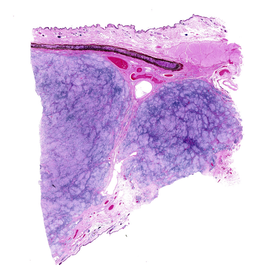
A 2.7 x 2.0 x 1.8 cm irregularly shaped section of haired skin diffusely expanded by an expansile cutaneous mass composed of several raised alopecic nodules labeled as removed from the left pinna is submitted for histopathologic examination.
Laboratory results:
N/A
Microscopic description:
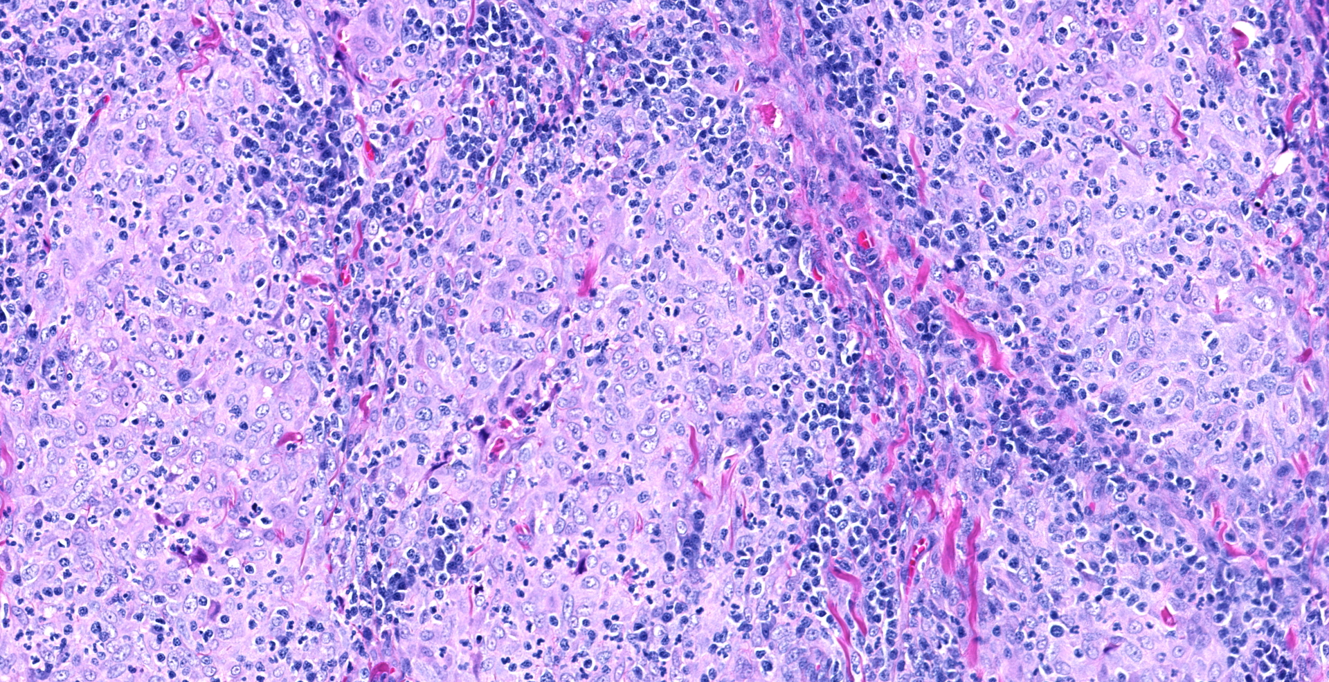
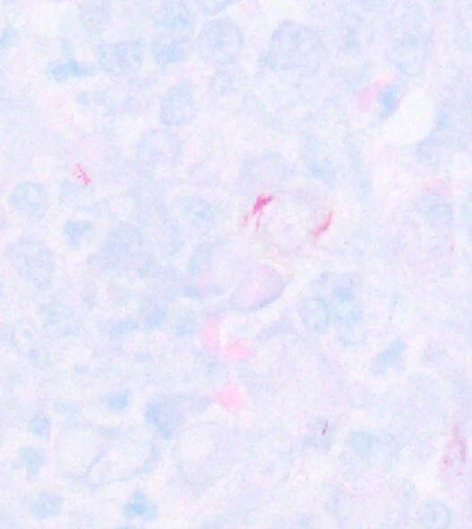
Haired skin left pinna cutaneous mass: The dermis is infiltrated, expanded, and effaced by marked pyogranulomatous inflammation composed of anastomosing nodular pyogranulomas. The inflammation is composed of large numbers of epithelioid macrophages admixed with smaller numbers of multinucleate giant cells, neutrophils, lymphocytes, and plasma cells. Neutrophils are often located centrally and the lymphocytes and plasma cells along the periphery of the pyogranulomatous nodules. Lymphoplasmacytic cuffs are often also seen around intralesional and perilesional blood vessels. Acid fast and modified acid-fast stains reveal acid-fast bacteria within moderate numbers of individually scattered macrophages; some affected macrophages only have 1 or 2 intracytoplasmic acid-fast bacteria while others contain numerous (>20+) acid-fast bacteria. The acid-fast bacteria are pleomorphic in appearance, ranging from coccoid to rod-shaped to beaded and filamentous. The inflammation extends on both sides of the pinnal cartilage and infiltrates the superficial dermis approaching (but not obscuring) the dermo-epidermal junction. The overlying epidermis is locally extensively thickened due to acanthosis and is covered by small amounts of compact hyperkeratotic to orthokeratotic keratin, with one superficially eroded area with local serohemorrhagic to neutrophilic exudation and crusting.
Contributor's morphologic diagnosis:
Haired skin left pinna: Severe, chronic, locally extensive, pyogranulomatous pinnal dermatitis, with intrahistiocytic acid-fast pleomorphic bacteria (canine leproid granuloma).
Contributor's comment:
Canine leproid granuloma (CLG) is an uncommon (but probably underdiagnosed) nodular mycobacterial disease of the skin.4,6 The condition usually occurs in short-coated breeds, with Boxer dogs and their crossbreeds remarkably overrepresented; Staffordshire Bull Terriers, Foxhounds, and Doberman Pinschers are also commonly affected. CLG presents clinically as single or multiple, firm, well-circumscribed nodules in the skin or subcutis that range in size from 2 mm to 5 cm; larger skin lesions may be alopecic or ulcerated.6 The lesions may be non-pruritic and painless, but mild signs of pain or pruritus have been reported in some animals.2 The dorsal surface of the pinna is a strongly predisposed site.6 In one large study, the head (including the ears) was affected in 85% of dogs, and the ears were involved in 64%; less commonly the distal extremities (especially the forelegs) may be affected.6 The great majority of CLG lesions are self-limiting with spontaneous regression of lesions often occurring within 3-6 months.4,6 Persistence of lesions beyond this time frame may warrant antibiotic treatment; cell-mediated immunity may be compromised in these patients and thus spontaneous regression may not occur.6 Extension of the infection to lymph nodes or visceral organs has not been reported.4,6
Cytologically, microscopic examination of fine needle aspirates of CLG syndrome lesions commonly reveals numerous, often spindle-shaped, macrophages admixed with variable numbers of lymphocytes and plasma cells, and fewer neutrophils. Usually few to moderate numbers of medium-length bacilli were detected within macrophages or extracellularly.1 Histologically, canine leproid granulomas are composed of nodular to diffuse, granulomatous to pyogranulomatous inflammation. Very small, or moderate numbers of acid-fast pleomorphic bacteria are present within small clusters of intralesional macrophages or are within individually scattered macrophages.1 The bacteria are acid-fast when stained with Ziehl-Neelsen stain and are usually bacilli when viewed in cytologic samples, but the microscopic appearance of the mycobacteria in histologic samples has been reported to be more pleomorphic and may include long, slender filaments in parallel sheaves, short and variably beaded bacilli, and highly beaded to coccoid forms.1
The causal agent of CLG is fastidious mycobacterial species which does not grow on synthetic mycobacterial media using standard microbiological methods. The disease has been reported in dogs from southern Africa, Australia, New Zealand, Brazil, and the United States (California, Florida, and the northeastern US).3,7 Recent publications using molecular techniques have shown genetic uniformity of the mycobacteria found in leproid granulomas from dogs in Australia, Brazil, USA, New Zealand, and Australia.2,3,5,8 The closest relative of the causative species of mycobacteria has been suggested to be Mycobacterium simiae.5 The mode(s) of transmission of the causative organism remains unknown; percutaneous inoculation has been suggested as a likely route of entry and biting insects have been proposed as potential mechanical vectors.2,6,8
Contributing Institution:
Atlantic Veterinary College
University of Prince Edward Island
JPC diagnosis:
Haired skin, pinna, ear: Dermatitis, nodular and granulomatous, focally extensive, severe.
JPC comment:
The contributor summarizes the sparse literature on this entity. Other differentials that may be considered include histiocytoma, plasmacytoma, fungal dermatitis, sterile granuloma, and sterile pyogranuloma syndrome.4
The hallmarks of histiocytoma, including epithelial hyperplasia, reniform nuclei, moderate mitotic rate, and top-heavy distribution were not present in this case. Plasmacytomas most often have binucleated cells, "helmet" cells, with well differentiated and distinguishable plasma cells at the periphery of the neoplasm. While a fungal infection could result in a similar lesion, the performed GMS stain revealed no fungal hyphae or yeasts. Sterile pyogranuloma syndrome also affects Boxers disproportionately but tend to have a vertical orientation that tracks hair follicles and have a more varied inflammatory cell content. In this case, the acid-fast bacteria provide the etiology for these lesions.
References:
- Charles J, Martin P, Wigney DI, Malik R, Love DN. Cytology and histopathology of canine leproid granuloma syndrome. Aust Vet J. 1999; 77(12):799-803.
- Conceição LG, Acha LM, Borges AS, Assis FG, Loures FH, e Silva FF. Epidemiology, clinical signs, histopathology and molecular characterization of canine leproid granuloma: a retrospective study of cases from Brazil. Vet Derm. 2011; 22(3):249-56.
- Foley JE, Borjesson D, l Gross T, Rand C, Needham M, Poland A. Clinical, microscopic, and molecular aspects of canine leproid granuloma in the United States. Vet Path. 2002; 39(2):234-9.
- Gross TL, Ihrke PJ, Walder EJ, Affolter VK. Skin diseases of the dog and cat: clinical and histopathologic diagnosis, 2nd ed. pp. 281-283. Blackwell Publishing, Ames, IA, 2005.
- Hughes MS, James G, Ball N, Scally M, Malik R, Wigney DI, Martin P, Chen S, Mitchell D, Love DN. Identification by 16S rRNA gene analyses of a potential novel mycobacterial species as an etiological agent of canine leproid granuloma syndrome. J Clin Microbiol. 2000; 38(3):953-9.
- Malik R, Smits B, Reppas G, Laprie C, O'brien C, Fyfe J. Ulcerated and nonulcerated nontuberculous cutaneous mycobacterial granulomas in cats and dogs. Vet Derm. 2013; 24(1):146-e33.
- Mauldin EA, Goldschmidt MH, Rankin SC. ISVD?5 Canine leproid granuloma in the northeastern United States. Vet Derm. 2004; 15:71-71.
- Smits B, Willis R, Malik R, Studdert V, Collins DM, Kawakami P, Graham D, Fyfe JA. Case clusters of leproid granulomas in foxhounds in New Zealand and Australia. Vet Derm. 2012; 23(6):465-e88.